3HE6 | pdb_00003he6

Crystal structure of mouse CD1d-alpha-galactosylceramide with mouse Valpha14-Vbeta8.2 NKT TCR


Experimental Data Snapshot

  • Method: X-RAY DIFFRACTION
  • Resolution: 2.90 Å
  • R-Value Free: 
    0.298 (Depositor), 0.310 (DCC) 
  • R-Value Work: 
    0.234 (Depositor), 0.265 (DCC) 
  • R-Value Observed: 
    0.237 (Depositor) 

Starting Models: experimental
View more details

wwPDB Validation 3D Report Full Report

Validation slider image for 3HE6

Ligand Structure Quality Assessment 


This is version 2.2 of the entry. See complete history

Literature

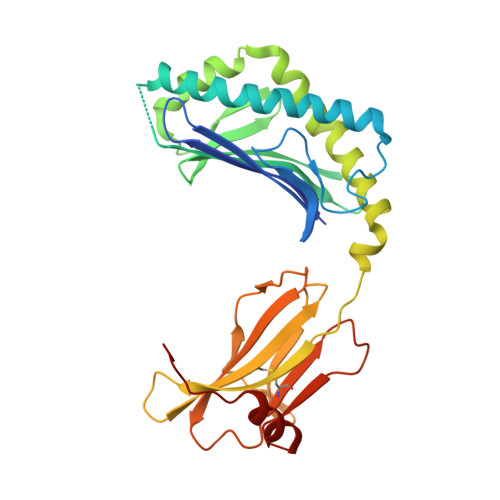
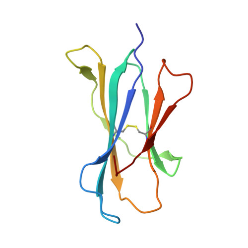
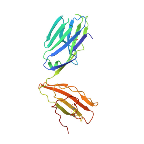
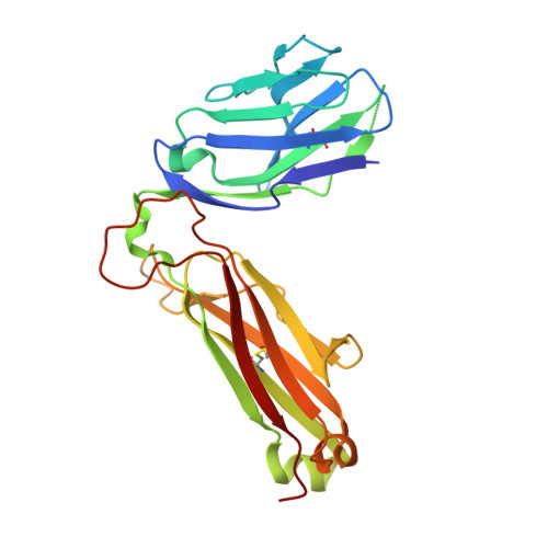
Differential recognition of CD1d-alpha-galactosyl ceramide by the V beta 8.2 and V beta 7 semi-invariant NKT T cell receptors

Pellicci, D.G.Patel, O.Kjer-Nielsen, L.Pang, S.S.Sullivan, L.C.Kyparissoudis, K.Brooks, A.G.Reid, H.H.Gras, S.Lucet, I.S.Koh, R.Smyth, M.J.Mallevaey, T.Matsuda, J.L.Gapin, L.McCluskey, J.Godfrey, D.I.Rossjohn, J.

(2009) Immunity 31: 47-59

  • DOI: https://doi.org/10.1016/j.immuni.2009.04.018
  • Primary Citation Related Structures: 
    3HE6, 3HE7, 3HUJ

  • PubMed Abstract: 

    The semi-invariant natural killer T cell receptor (NKT TCR) recognizes CD1d-lipid antigens. Although the TCR alpha chain is typically invariant, the beta chain expression is more diverse, where three V beta chains are commonly expressed in mice. We report the structures of V alpha 14-V beta 8.2 and V alpha 14-V beta 7 NKT TCRs in complex with CD1d-alpha-galactosylceramide (alpha-GalCer) and the 2.5 A structure of the human NKT TCR-CD1d-alpha-GalCer complex. Both V beta 8.2 and V beta 7 NKT TCRs and the human NKT TCR ligated CD1d-alpha-GalCer in a similar manner, highlighting the evolutionarily conserved interaction. However, differences within the V beta domains of the V beta 8.2 and V beta 7 NKT TCR-CD1d complexes resulted in altered TCR beta-CD1d-mediated contacts and modulated recognition mediated by the invariant alpha chain. Mutagenesis studies revealed the differing contributions of V beta 8.2 and V beta 7 residues within the CDR2 beta loop in mediating contacts with CD1d. Collectively we provide a structural basis for the differential NKT TCR V beta usage in NKT cells.


  • Organizational Affiliation
    • Department of Microbiology and Immunology, University of Melbourne, Parkville, Victoria 3010, Australia.

Macromolecule Content 

  • Total Structure Weight: 97.99 kDa 
  • Atom Count: 6,703 
  • Modeled Residue Count: 824 
  • Deposited Residue Count: 852 
  • Unique protein chains: 4

Macromolecules

Find similar proteins by:|  3D Structure
Entity ID: 1
MoleculeChains  Sequence LengthOrganismDetailsImage
Antigen-presenting glycoprotein CD1d1302Mus musculusMutation(s): 0 
Gene Names: Cd1d1Cd1.1
UniProt & NIH Common Fund Data Resources
Find proteins for P11609 (Mus musculus)
Explore P11609 
Go to UniProtKB:  P11609
IMPC:  MGI:107674
Entity Groups
Sequence Clusters30% Identity50% Identity70% Identity90% Identity95% Identity100% Identity
UniProt GroupP11609
Glycosylation
Glycosylation Sites: 3Go to GlyGen: P11609-1
Sequence Annotations
Expand
Reference Sequence
Find similar proteins by:|  3D Structure
Entity ID: 2
MoleculeChains  Sequence LengthOrganismDetailsImage
Beta-2-microglobulin99Mus musculusMutation(s): 0 
Gene Names: B2m
UniProt & NIH Common Fund Data Resources
Find proteins for P01887 (Mus musculus)
Explore P01887 
Go to UniProtKB:  P01887
IMPC:  MGI:88127
Entity Groups
Sequence Clusters30% Identity50% Identity70% Identity90% Identity95% Identity100% Identity
UniProt GroupP01887
Sequence Annotations
Expand
Reference Sequence
Find similar proteins by:|  3D Structure
Entity ID: 3
MoleculeChains  Sequence LengthOrganismDetailsImage
Valpha14(mouse variable domain, human constant domain)207Mus musculusMutation(s): 0 
Find similar proteins by:|  3D Structure
Entity ID: 4
MoleculeChains  Sequence LengthOrganismDetailsImage
Vbeta8.2(mouse variable domain, human constant domain)244Mus musculusMutation(s): 0 
UniProt
Find proteins for P01850 (Homo sapiens)
Explore P01850 
Go to UniProtKB:  P01850
Entity Groups
Sequence Clusters30% Identity50% Identity70% Identity90% Identity95% Identity100% Identity
UniProt GroupP01850
Sequence Annotations
Expand
Reference Sequence

Oligosaccharides

Help  
Entity ID: 5
MoleculeChains Length2D Diagram GlycosylationD Interactions
2-acetamido-2-deoxy-beta-D-glucopyranose-(1-4)-2-acetamido-2-deoxy-beta-D-glucopyranose
E
2N-Glycosylation
Glycosylation Resources
GlyTouCan: G42666HT
GlyCosmos: G42666HT
GlyGen: G42666HT

Small Molecules

Ligands 2 Unique
IDChains Name / Formula / InChI Key2D Diagram3D Interactions
AGH

Query on AGH


Download:Ideal Coordinates CCD File
H [auth A]N-{(1S,2R,3S)-1-[(ALPHA-D-GALACTOPYRANOSYLOXY)METHYL]-2,3-DIHYDROXYHEPTADECYL}HEXACOSANAMIDE
C50 H99 N O9
VQFKFAKEUMHBLV-BYSUZVQFSA-N
NAG

Query on NAG


Download:Ideal Coordinates CCD File
F [auth A],
G [auth A]
2-acetamido-2-deoxy-beta-D-glucopyranose
C8 H15 N O6
OVRNDRQMDRJTHS-FMDGEEDCSA-N

Experimental Data & Validation

Experimental Data

  • Method: X-RAY DIFFRACTION
  • Resolution: 2.90 Å
  • R-Value Free:  0.298 (Depositor), 0.310 (DCC) 
  • R-Value Work:  0.234 (Depositor), 0.265 (DCC) 
  • R-Value Observed: 0.237 (Depositor) 
Space Group: P 21 21 21
Unit Cell:
Length ( Å )Angle ( ˚ )
a = 61.113α = 90
b = 81.506β = 90
c = 233.451γ = 90
Software Package:
Software NamePurpose
ADSCdata collection
PHASERphasing
REFMACrefinement
MOSFLMdata reduction
SCALAdata scaling

Structure Validation

View Full Validation Report


Ligand Structure Quality Assessment 


Entry History 

Deposition Data

Revision History  (Full details and data files)

  • Version 1.0: 2009-07-28
    Type: Initial release
  • Version 1.1: 2011-07-13
    Changes: Non-polymer description, Version format compliance
  • Version 1.2: 2020-01-29
    Changes: Advisory, Database references, Derived calculations, Source and taxonomy, Structure summary
  • Version 2.0: 2020-07-29
    Type: Remediation
    Reason: Carbohydrate remediation
    Changes: Atomic model, Data collection, Derived calculations, Structure summary
  • Version 2.1: 2023-11-01
    Changes: Data collection, Database references, Refinement description, Structure summary
  • Version 2.2: 2024-10-30
    Changes: Structure summary