3HBR | pdb_00003hbr

Crystal structure of OXA-48 beta-lactamase


Experimental Data Snapshot

  • Method: X-RAY DIFFRACTION
  • Resolution: 1.90 Å
  • R-Value Free: 
    0.286 (Depositor), 0.286 (DCC) 
  • R-Value Work: 
    0.217 (Depositor), 0.217 (DCC) 
  • R-Value Observed: 
    0.223 (Depositor) 

Starting Model: experimental
View more details

wwPDB Validation 3D Report Full Report

Validation slider image for 3HBR

This is version 1.3 of the entry. See complete history

Literature

Crystal structure of the OXA-48 beta-lactamase reveals mechanistic diversity among class D carbapenemases.

Docquier, J.D.Calderone, V.De Luca, F.Benvenuti, M.Giuliani, F.Bellucci, L.Tafi, A.Nordmann, P.Botta, M.Rossolini, G.M.Mangani, S.

(2009) Chem Biol 16: 540-547

  • DOI: https://doi.org/10.1016/j.chembiol.2009.04.010
  • Primary Citation Related Structures: 
    3HBR

  • PubMed Abstract: 

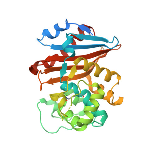
    Carbapenem-hydrolyzing class D beta-lactamases (CHDLs) are enzymes found in important Gram-negative pathogens (mainly Acinetobacter baumannii and Enterobacteriaceae) that confer resistance to beta-lactam antibiotics, and notably carbapenems. The crystal structure of the OXA-48 carbapenemase was determined at pH 7.5 and at a resolution of 1.9 A. Surprisingly, and by contrast with OXA-24, the only other CHDL of known crystal structure, the structure of OXA-48 was similar to OXA-10, an enzyme devoid of carbapenemase activity, indicating that the hydrolysis of these compounds could depend on subtle changes in the active site region. Moreover, the active site groove of OXA-48 was different from that of OXA-24 in shape, dimensions, and charge distribution. Molecular dynamics pointed to the functional relevance of residues located in or close to the beta5-beta6 loop and allowed us to propose a mechanism for carbapenem hydrolysis by OXA-48.


  • Organizational Affiliation
    • Dipartimento di Biologia Molecolare, Università di Siena, Italy.

Macromolecule Content 

  • Total Structure Weight: 122.01 kDa 
  • Atom Count: 8,635 
  • Modeled Residue Count: 960 
  • Deposited Residue Count: 1,060 
  • Unique protein chains: 1

Macromolecules

Find similar proteins by:|  3D Structure
Entity ID: 1
MoleculeChains  Sequence LengthOrganismDetailsImage
OXA-48
A, B, C, D
265Klebsiella pneumoniaeMutation(s): 0 
Gene Names: bla OXA-48
EC: 3.5.2.6
UniProt
Find proteins for Q6XEC0 (Klebsiella pneumoniae)
Explore Q6XEC0 
Go to UniProtKB:  Q6XEC0
Entity Groups
Sequence Clusters30% Identity50% Identity70% Identity90% Identity95% Identity100% Identity
UniProt GroupQ6XEC0
Sequence Annotations
Expand
Reference Sequence

Experimental Data & Validation

Experimental Data

  • Method: X-RAY DIFFRACTION
  • Resolution: 1.90 Å
  • R-Value Free:  0.286 (Depositor), 0.286 (DCC) 
  • R-Value Work:  0.217 (Depositor), 0.217 (DCC) 
  • R-Value Observed: 0.223 (Depositor) 
Space Group: P 1 21 1
Unit Cell:
Length ( Å )Angle ( ˚ )
a = 63.701α = 90
b = 107.185β = 111.04
c = 80.787γ = 90
Software Package:
Software NamePurpose
ADSCdata collection
MOLREPphasing
REFMACrefinement
MOSFLMdata reduction
SCALAdata scaling

Structure Validation

View Full Validation Report


Entry History 

Deposition Data

Revision History  (Full details and data files)

  • Version 1.0: 2009-06-23
    Type: Initial release
  • Version 1.1: 2011-07-13
    Changes: Version format compliance
  • Version 1.2: 2023-09-06
    Changes: Data collection, Database references, Derived calculations, Refinement description
  • Version 1.3: 2023-11-22
    Changes: Data collection