Crystal structure of ED-Eya2: insight into dual roles as a protein tyrosine phosphatase and a transcription factor
Jung, S.K., Jeong, D.G., Chung, S.J., Kim, J.H., Park, B.C., Tonks, N.K., Ryu, S.E., Kim, S.J.(2010) FASEB J 24: 560-569
- PubMed: 19858093
- DOI: https://doi.org/10.1096/fj.09-143891
- Primary Citation of Related Structures:
3HB0, 3HB1 - PubMed Abstract:
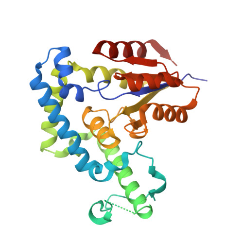
Eya proteins are transcription factors that play pivotal roles in organ formation during development by mediating interactions between Sine Oculis (SO) and Dachshund (DAC). Remarkably, the transcriptional activity of Eya proteins is regulated by a dephosphorylating activity within its Eya domain (ED). However, the molecular basis for the link between catalytic and transcriptional activities remains unclear. Here we report the first description of the crystal structure of the ED of human Eya2 (ED-Eya2), determined at 2.4-A resolution. In stark contrast to other members of the haloacid dehalogenase (HAD) family to which ED-Eya2 belongs, the helix-bundle motif (HBM) is elongated along the back of the catalytic site. This not only results in a structure that accommodates large protein substrates but also positions the catalytic and the SO-interacting sites on opposite faces, which suggests that SO binding is not directly affected by catalytic function. Based on the observation that the DAC-binding site is located between the catalytic core and SO binding sites within ED-Eya2, we propose that catalytic activity can be translated to SO binding through DAC, which acts as a transcriptional switch. We also captured at two stages of reaction cycles-acyl-phosphate intermediate and transition state of hydrolysis step, which provided a detailed view of reaction mechanism. The ED-Eya2 structure defined here serves as a model for other members of the Eya family and provides a framework for understanding the role of Eya phosphatase mutations in disease.
Organizational Affiliation:
Medical Proteomics Research Center, Korea Research Institute of Bioscience and Biotechnology, 52 Eoeun-Dong, Yuseong-Gu, Daejeon, 305-600, Korea.


