3HAN | pdb_00003han

Crystal structure of bacteriorhodopsin mutant V49A crystallized from bicelles


Experimental Data Snapshot

  • Method: X-RAY DIFFRACTION
  • Resolution: 2.75 Å
  • R-Value Free: 
    0.282 (Depositor), 0.279 (DCC) 
  • R-Value Work: 
    0.222 (Depositor), 0.219 (DCC) 
  • R-Value Observed: 
    0.227 (Depositor) 

wwPDB Validation 3D Report Full Report

Validation slider image for 3HAN

Ligand Structure Quality Assessment 


This is version 1.4 of the entry. See complete history

Literature

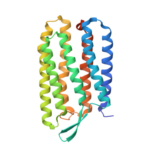
Similar energetic contributions of packing in the core of membrane and water-soluble proteins.

Joh, N.H.Oberai, A.Yang, D.Whitelegge, J.P.Bowie, J.U.

(2009) J Am Chem Soc 131: 10846-10847

  • DOI: https://doi.org/10.1021/ja904711k
  • Primary Citation Related Structures: 
    3HAN, 3HAO, 3HAP, 3HAQ, 3HAR, 3HAS

  • PubMed Abstract: 

    A major driving force for water-soluble protein folding is the hydrophobic effect, but membrane proteins cannot make use of this stabilizing contribution in the apolar core of the bilayer. It has been proposed that membrane proteins compensate by packing more efficiently. We therefore investigated packing contributions experimentally by observing the energetic and structural consequences of cavity creating mutations in the core of a membrane protein. We observed little difference in the packing energetics of water and membrane soluble proteins. Our results imply that other mechanisms are employed to stabilize the structure of membrane proteins.


  • Organizational Affiliation
    • Department of Chemistry and Biochemistry, UCLA-DOE Center for Genomics and Proteomics, Molecular Biology Institute, University of California, Los Angeles, California 90095, USA.

Macromolecule Content 

  • Total Structure Weight: 27.19 kDa 
  • Atom Count: 1,771 
  • Modeled Residue Count: 225 
  • Deposited Residue Count: 249 
  • Unique protein chains: 1

Macromolecules

Find similar proteins by:|  3D Structure
Entity ID: 1
MoleculeChains  Sequence LengthOrganismDetailsImage
Bacteriorhodopsin249Halobacterium salinarumMutation(s): 1 
Gene Names: bopVNG_1467G
Membrane Entity: Yes 
UniProt
Find proteins for P02945 (Halobacterium salinarum (strain ATCC 700922 / JCM 11081 / NRC-1))
Explore P02945 
Go to UniProtKB:  P02945
Entity Groups
Sequence Clusters30% Identity50% Identity70% Identity90% Identity95% Identity100% Identity
UniProt GroupP02945
Sequence Annotations
Expand
Reference Sequence

Small Molecules

Ligands 1 Unique
IDChains Name / Formula / InChI Key2D Diagram3D Interactions
RET

Query on RET


Download:Ideal Coordinates CCD File
B [auth A]RETINAL
C20 H28 O
NCYCYZXNIZJOKI-OVSJKPMPSA-N

Experimental Data & Validation

Experimental Data

  • Method: X-RAY DIFFRACTION
  • Resolution: 2.75 Å
  • R-Value Free:  0.282 (Depositor), 0.279 (DCC) 
  • R-Value Work:  0.222 (Depositor), 0.219 (DCC) 
  • R-Value Observed: 0.227 (Depositor) 
Space Group: C 2 2 21
Unit Cell:
Length ( Å )Angle ( ˚ )
a = 44.948α = 90
b = 102.878β = 90
c = 128.422γ = 90
Software Package:
Software NamePurpose
DENZOdata reduction
SCALEPACKdata scaling
REFMACrefinement
PDB_EXTRACTdata extraction

Structure Validation

View Full Validation Report


Ligand Structure Quality Assessment 


Entry History 

Deposition Data

Revision History  (Full details and data files)

  • Version 1.0: 2009-09-22
    Type: Initial release
  • Version 1.1: 2011-07-13
    Changes: Version format compliance
  • Version 1.2: 2017-11-01
    Changes: Refinement description
  • Version 1.3: 2021-10-13
    Changes: Database references, Derived calculations
  • Version 1.4: 2024-11-06
    Changes: Data collection, Structure summary