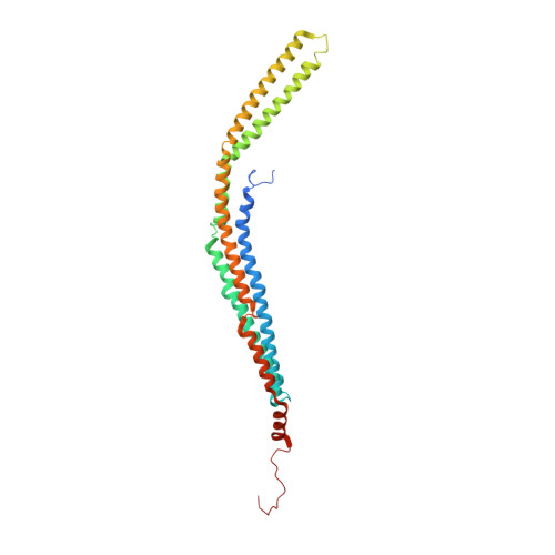
3HAI

Crystal structure of human PACSIN1 F-BAR domain (P21 lattice)


Experimental Data Snapshot

  • Method: X-RAY DIFFRACTION
  • Resolution: 2.88 Å
  • R-Value Free: 0.319 
  • R-Value Work: 0.282 
  • R-Value Observed: 0.284 

wwPDB Validation   3D Report Full Report


This is version 1.2 of the entry. See complete history


Literature

Molecular mechanism of membrane constriction and tubulation mediated by the F-BAR protein Pacsin/Syndapin.

Wang, Q.Navarro, M.V.Peng, G.Molinelli, E.Lin Goh, S.Judson, B.L.Rajashankar, K.R.Sondermann, H.

(2009) Proc Natl Acad Sci U S A 106: 12700-12705

  • DOI: https://doi.org/10.1073/pnas.0902974106
  • Primary Citation of Related Structures:  
    3HAH, 3HAI, 3HAJ

  • PubMed Abstract: 

    Peripheral membrane proteins of the Bin/amphiphysin/Rvs (BAR) and Fer-CIP4 homology-BAR (F-BAR) family participate in cellular membrane trafficking and have been shown to generate membrane tubules. The degree of membrane bending appears to be encoded in the structure and immanent curvature of the particular protein domains, with BAR and F-BAR domains inducing high- and low-curvature tubules, respectively. In addition, oligomerization and the formation of ordered arrays influences tubule stabilization. Here, the F-BAR domain-containing protein Pacsin was found to possess a unique activity, creating small tubules and tubule constrictions, in addition to the wide tubules characteristic for this subfamily. Based on crystal structures of the F-BAR domain of Pacsin and mutagenesis studies, vesiculation could be linked to the presence of unique structural features distinguishing it from other F-BAR proteins. Tubulation was suppressed in the context of the full-length protein, suggesting that Pacsin is autoinhibited in solution. The regulated deformation of membranes and promotion of tubule constrictions by Pacsin suggests a more versatile function of these proteins in vesiculation and endocytosis beyond their role as scaffold proteins.


  • Organizational Affiliation

    Department of Molecular Medicine, College of Veterinary Medicine, Cornell University, Ithaca, NY 14853, USA.


Macromolecules
Find similar proteins by:  (by identity cutoff)  |  3D Structure
Entity ID: 1
MoleculeChains Sequence LengthOrganismDetailsImage
human PACSIN1 F-BAR
A, B, C, D
308Homo sapiensMutation(s): 0 
Gene Names: PACSIN1KIAA1379
UniProt & NIH Common Fund Data Resources
Find proteins for Q9BY11 (Homo sapiens)
Explore Q9BY11 
Go to UniProtKB:  Q9BY11
PHAROS:  Q9BY11
GTEx:  ENSG00000124507 
Entity Groups  
Sequence Clusters30% Identity50% Identity70% Identity90% Identity95% Identity100% Identity
UniProt GroupQ9BY11
Sequence Annotations
Expand
  • Reference Sequence
Small Molecules
Ligands 1 Unique
IDChains Name / Formula / InChI Key2D Diagram3D Interactions
CA
Query on CA

Download Ideal Coordinates CCD File 
E [auth A]CALCIUM ION
Ca
BHPQYMZQTOCNFJ-UHFFFAOYSA-N
Experimental Data & Validation

Experimental Data

  • Method: X-RAY DIFFRACTION
  • Resolution: 2.88 Å
  • R-Value Free: 0.319 
  • R-Value Work: 0.282 
  • R-Value Observed: 0.284 
  • Space Group: P 1 21 1
Unit Cell:
Length ( Å )Angle ( ˚ )
a = 163.62α = 90
b = 32.16β = 111.23
c = 181.16γ = 90
Software Package:
Software NamePurpose
ADSCdata collection
MLPHAREphasing
PHENIXrefinement
HKL-2000data reduction
HKL-2000data scaling

Structure Validation

View Full Validation Report


Entry History 

Deposition Data

Revision History  (Full details and data files)

  • Version 1.0: 2009-06-16
    Type: Initial release
  • Version 1.1: 2011-07-13
    Changes: Version format compliance
  • Version 1.2: 2024-02-21
    Changes: Data collection, Database references, Derived calculations