3H8Y | pdb_00003h8y

Crystal structure of carboxysome small shell protein CsoS1C from Halothiobacillus neapolitanus


Experimental Data Snapshot

  • Method: X-RAY DIFFRACTION
  • Resolution: 2.51 Å
  • R-Value Free: 
    0.311 (Depositor), 0.310 (DCC) 
  • R-Value Work: 
    0.255 (Depositor), 0.255 (DCC) 
  • R-Value Observed: 
    0.258 (Depositor) 

Starting Model: experimental
View more details

wwPDB Validation 3D Report Full Report

Validation slider image for 3H8Y

This is version 1.3 of the entry. See complete history

Literature

Analysis of lattice-translocation disorder in the layered hexagonal structure of carboxysome shell protein CsoS1C

Tsai, Y.Sawaya, M.R.Yeates, T.O.

(2009) Acta Crystallogr D Biol Crystallogr 65: 980-988

  • DOI: https://doi.org/10.1107/S0907444909025153
  • Primary Citation Related Structures: 
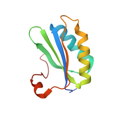
    3H8Y

  • PubMed Abstract: 

    Lattice-translocation or crystal order-disorder phenomena occur when some layers or groups of molecules in a crystal are randomly displaced relative to other groups of molecules by a discrete set of vectors. In previous work, the effects of lattice translocation on diffraction intensities have been corrected by considering that the observed intensities are the product of the intensities from an ideal crystal (lacking disorder) multiplied by the squared magnitude of the Fourier transform of the set of translocation vectors. Here, the structure determination is presented of carboxysome protein CsoS1C from Halothiobacillius neapolitanus in a crystal exhibiting a lattice translocation with unique features. The diffraction data are fully accounted for by a crystal unit cell composed of two layers of cyclic protein hexamers. The first layer is fully ordered (i.e. has one fixed position), while the second layer randomly takes one of three alternative positions whose displacements are related to each other by threefold symmetry. Remarkably, the highest symmetry present in the crystal is P3, yet the intensity data (and the Patterson map) obey 6/m instead of \overline 3 symmetry; the intensities exceed the symmetry expected from combining the crystal space group with an inversion center. The origin of this rare phenomenon, known as symmetry enhancement, is discussed and shown to be possible even for a perfectly ordered crystal. The lattice-translocation treatment described here may be useful in analyzing other cases of disorder in which layers or groups of molecules are shifted in multiple symmetry-related directions.


  • Organizational Affiliation
    • Molecular Biology Institute, University of California Los Angeles, Los Angeles, CA, USA.

Macromolecule Content 

  • Total Structure Weight: 9.93 kDa 
  • Atom Count: 699 
  • Modeled Residue Count: 95 
  • Deposited Residue Count: 98 
  • Unique protein chains: 1

Macromolecules

Find similar proteins by:|  3D Structure
Entity ID: 1
MoleculeChains  Sequence LengthOrganismDetailsImage
Major carboxysome shell protein 1C98Halothiobacillus neapolitanusMutation(s): 0 
Gene Names: csoS1C
UniProt
Find proteins for P45688 (Halothiobacillus neapolitanus (strain ATCC 23641 / DSM 15147 / CIP 104769 / NCIMB 8539 / c2))
Explore P45688 
Go to UniProtKB:  P45688
Entity Groups
Sequence Clusters30% Identity50% Identity70% Identity90% Identity95% Identity100% Identity
UniProt GroupP45688
Sequence Annotations
Expand
Reference Sequence

Experimental Data & Validation

Experimental Data

  • Method: X-RAY DIFFRACTION
  • Resolution: 2.51 Å
  • R-Value Free:  0.311 (Depositor), 0.310 (DCC) 
  • R-Value Work:  0.255 (Depositor), 0.255 (DCC) 
  • R-Value Observed: 0.258 (Depositor) 
Space Group: P 6
Unit Cell:
Length ( Å )Angle ( ˚ )
a = 66.675α = 90
b = 66.675β = 90
c = 63.007γ = 120
Software Package:
Software NamePurpose
DENZOdata reduction
SCALEPACKdata scaling
PHASERphasing
REFMACrefinement
PDB_EXTRACTdata extraction
ADSCdata collection

Structure Validation

View Full Validation Report


Entry History 

Deposition Data

Revision History  (Full details and data files)

  • Version 1.0: 2009-09-01
    Type: Initial release
  • Version 1.1: 2011-07-13
    Changes: Version format compliance
  • Version 1.2: 2017-11-01
    Changes: Refinement description
  • Version 1.3: 2023-09-06
    Changes: Data collection, Database references, Refinement description