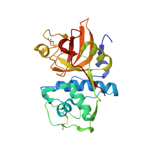
3H8B

A combined crystallographic and molecular dynamics study of cathepsin-L retro-binding inhibitors(compound 9)


Experimental Data Snapshot

  • Method: X-RAY DIFFRACTION
  • Resolution: 1.80 Å
  • R-Value Free: 0.250 
  • R-Value Work: 0.216 

wwPDB Validation   3D Report Full Report


Ligand Structure Quality Assessment 


This is version 1.3 of the entry. See complete history


Literature

A combined crystallographic and molecular dynamics study of cathepsin L retrobinding inhibitors

Shenoy, R.T.Chowdhury, S.F.Kumar, S.Joseph, L.Purisima, E.O.Sivaraman, J.

(2009) J Med Chem 52: 6335-6346

  • DOI: https://doi.org/10.1021/jm900596y
  • Primary Citation of Related Structures:  
    3H89, 3H8B, 3H8C

  • PubMed Abstract: 

    We report the crystal structures of three noncovalent retrobinding inhibitors in complex with mature cathepsin L up to resolutions of 2.5, 1.8, and 2.5 A, respectively. These inhibitors were Bpa-(Nepsilon-Bpa)Lys-DArg-Tyr-Npe, Bpa-(Nepsilon-Bpa)Lys-DArg-Phe-Npe, and Bpa-MCys-DArg-Phe-Npe, where Bpa = biphenylacetyl and Pea = N-phenylethyl. These were selected to clarify the binding mode of the biphenyl groups in the S' subsites because the addition of a second biphenyl does not improve potency. Examination of the symmetry-related monomers in the crystal structures revealed inhibitor-inhibitor crystal packing interactions. Molecular dynamics simulations were then used to explore the structure and dynamical behavior of the isolated protein-ligand complexes in solution. In the simulations, the backbone biphenyl groups for all three inhibitors ended up in the same location despite having started out in different orientations in the initial crystal structure conformations. The lack of improved potency of the larger inhibitors over the smaller one is attributed to a correspondingly greater entropic cost of binding.


  • Organizational Affiliation

    Department of Biological Sciences, National University of Singapore, 14 Science Drive 4, Singapore 117543.


Macromolecules
Find similar proteins by:  (by identity cutoff)  |  3D Structure
Entity ID: 1
MoleculeChains Sequence LengthOrganismDetailsImage
Cathepsin L1
A, B, C, D, E
A, B, C, D, E, F
220Homo sapiensMutation(s): 0 
Gene Names: Cathepsin L
EC: 3.4.22.15
UniProt & NIH Common Fund Data Resources
Find proteins for P07711 (Homo sapiens)
Explore P07711 
Go to UniProtKB:  P07711
PHAROS:  P07711
GTEx:  ENSG00000135047 
Entity Groups  
Sequence Clusters30% Identity50% Identity70% Identity90% Identity95% Identity100% Identity
UniProt GroupP07711
Sequence Annotations
Expand
  • Reference Sequence
Experimental Data & Validation

Experimental Data

  • Method: X-RAY DIFFRACTION
  • Resolution: 1.80 Å
  • R-Value Free: 0.250 
  • R-Value Work: 0.216 
  • Space Group: C 1 2 1
Unit Cell:
Length ( Å )Angle ( ˚ )
a = 61.99α = 90
b = 99.234β = 90.05
c = 206.462γ = 90
Software Package:
Software NamePurpose
HKL-2000data collection
AMoREphasing
CNSrefinement
HKL-2000data reduction
HKL-2000data scaling

Structure Validation

View Full Validation Report


Ligand Structure Quality Assessment 


Entry History 

Deposition Data

Revision History  (Full details and data files)

  • Version 1.0: 2009-10-20
    Type: Initial release
  • Version 1.1: 2011-07-13
    Changes: Version format compliance
  • Version 1.2: 2014-02-05
    Changes: Database references
  • Version 1.3: 2024-11-20
    Changes: Data collection, Database references, Derived calculations, Structure summary