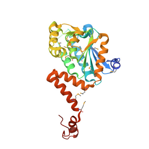
2.15 Angstrom Resolution Crystal Structure of Naphthoate Synthase from Salmonella typhimurium.
Minasov, G., Wawrzak, Z., Skarina, T., Onopriyenko, O., Peterson, S.N., Savchenko, A., Anderson, W.F., Center for Structural Genomics of Infectious Diseases (CSGID)To be published.