3GYO

Se-Met Rtt106p


Experimental Data Snapshot

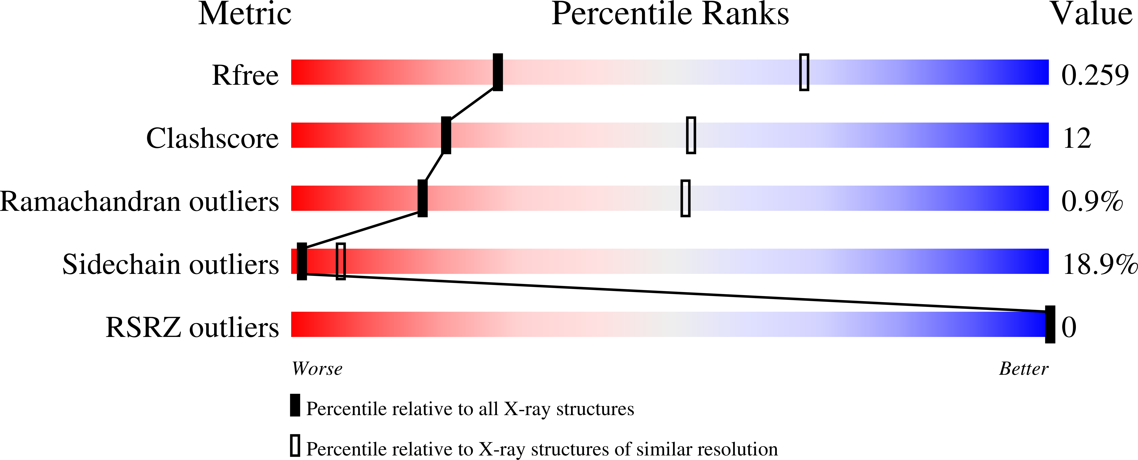
  • Method: X-RAY DIFFRACTION
  • Resolution: 3.10 Å
  • R-Value Free: 0.260 
  • R-Value Work: 0.215 
  • R-Value Observed: 0.217 

wwPDB Validation   3D Report Full Report


This is version 1.4 of the entry. See complete history


Literature

Structural analysis of Rtt106p reveals a DNA-binding role required for heterochromatin silencing

Liu, Y.Huang, H.Zhou, B.O.Wang, S.S.Hu, Y.Li, X.Liu, J.Zang, J.Niu, L.Wu, J.Zhou, J.Q.Teng, M.Shi, Y.

(2010) J Biol Chem 285: 4251-4262

  • DOI: https://doi.org/10.1074/jbc.M109.055996
  • Primary Citation of Related Structures:  
    3GYO, 3GYP

  • PubMed Abstract: 

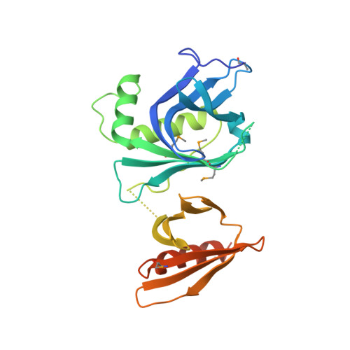
    Rtt106p is a Saccharomyces cerevisiae histone chaperone with roles in heterochromatin silencing and nucleosome assembly. The molecular mechanism by which Rtt106p engages in chromatin dynamics remains unclear. Here, we report the 2.5 A crystal structure of the core domain of Rtt106p, which adopts an unusual "double pleckstrin homology" domain architecture that represents a novel structural mode for histone chaperones. A histone H3-H4-binding region and a novel double-stranded DNA-binding region have been identified. Mutagenesis studies reveal that the histone and DNA binding activities of Rtt106p are involved in Sir protein-mediated heterochromatin formation. Our results uncover the structural basis of the diverse functions of Rtt106p and provide new insights into its cellular roles.


  • Organizational Affiliation

    From the Hefei National Laboratory for Physical Sciences at Microscale and School of Life Science, University of Science and Technology of China, Hefei, Anhui 230026 and.


Macromolecules
Find similar proteins by:  (by identity cutoff)  |  3D Structure
Entity ID: 1
MoleculeChains Sequence LengthOrganismDetailsImage
Histone chaperone RTT106261Saccharomyces cerevisiaeMutation(s): 1 
Gene Names: RTT106
UniProt
Find proteins for P40161 (Saccharomyces cerevisiae (strain ATCC 204508 / S288c))
Explore P40161 
Go to UniProtKB:  P40161
Entity Groups  
Sequence Clusters30% Identity50% Identity70% Identity90% Identity95% Identity100% Identity
UniProt GroupP40161
Sequence Annotations
Expand
  • Reference Sequence
Small Molecules
Modified Residues  1 Unique
IDChains TypeFormula2D DiagramParent
MSE
Query on MSE
A
L-PEPTIDE LINKINGC5 H11 N O2 SeMET
Experimental Data & Validation

Experimental Data

  • Method: X-RAY DIFFRACTION
  • Resolution: 3.10 Å
  • R-Value Free: 0.260 
  • R-Value Work: 0.215 
  • R-Value Observed: 0.217 
  • Space Group: F 4 3 2
Unit Cell:
Length ( Å )Angle ( ˚ )
a = 262.498α = 90
b = 262.498β = 90
c = 262.498γ = 90
Software Package:
Software NamePurpose
DENZOdata reduction
SCALEPACKdata scaling
SOLVEphasing
REFMACrefinement
PDB_EXTRACTdata extraction
HKL-2000data reduction

Structure Validation

View Full Validation Report


Entry History 

Deposition Data

Revision History  (Full details and data files)

  • Version 1.0: 2009-12-08
    Type: Initial release
  • Version 1.1: 2011-07-13
    Changes: Version format compliance
  • Version 1.2: 2017-11-01
    Changes: Refinement description
  • Version 1.3: 2021-11-10
    Changes: Database references, Derived calculations
  • Version 1.4: 2024-10-30
    Changes: Data collection, Structure summary