3GVN | pdb_00003gvn

The 1.2 Angstroem crystal structure of an E.coli tRNASer acceptor stem microhelix reveals two magnesium binding sites


Experimental Data Snapshot

  • Method: X-RAY DIFFRACTION
  • Resolution: 1.20 Å
  • R-Value Free: 
    0.201 (Depositor), 0.185 (DCC) 
  • R-Value Work: 
    0.190 (Depositor), 0.193 (DCC) 

Starting Model: experimental
View more details

wwPDB Validation 3D Report Full Report

Validation slider image for 3GVN

This is version 1.2 of the entry. See complete history

Literature

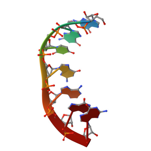
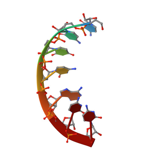
The 1.2A crystal structure of an E. coli tRNASer)acceptor stem microhelix reveals two magnesium binding sites.

Eichert, A.Furste, J.P.Schreiber, A.Perbandt, M.Betzel, C.Erdmann, V.A.Forster, C.

(2009) Biochem Biophys Res Commun 386: 368-373

  • DOI: https://doi.org/10.1016/j.bbrc.2009.06.048
  • Primary Citation Related Structures: 
    3GVN

  • PubMed Abstract: 

    tRNA identity elements assure the correct aminoacylation of tRNAs by the cognate aminoacyl-tRNA synthetases. tRNA(Ser) belongs to the so-called class II system, in which the identity elements are rather simple and are mostly located in the acceptor stem region, in contrast to 'class I', where tRNA determinants are more complex and are located within different regions of the tRNA. The structure of an Escherichia coli tRNA(Ser) acceptor stem microhelix was solved by high resolution X-ray structure analysis. The RNA crystallizes in the space group C2, with one molecule per asymmetric unit and with the cell constants a=35.79, b=39.13, c=31.37A, and beta=111.1 degrees . A defined hydration pattern of 97 water molecules surrounds the tRNA(Ser) acceptor stem microhelix. Additionally, two magnesium binding sites were detected in the tRNA(Ser) aminoacyl stem.


  • Organizational Affiliation
    • Institute of Chemistry and Biochemistry, Free University Berlin, Thielallee 63, 14195 Berlin, Germany.

Macromolecule Content 

  • Total Structure Weight: 4.48 kDa 
  • Atom Count: 392 
  • Modeled Residue Count: 14 
  • Deposited Residue Count: 14 
  • Unique nucleic acid chains: 2

Macromolecules

Find similar nucleic acids by:  Sequence
Entity ID: 1
MoleculeChains LengthOrganismImage
5'-R(*GP*GP*UP*GP*AP*GP*G)-3'7N/A
Sequence Annotations
Expand
Reference Sequence
Find similar nucleic acids by:  Sequence
Entity ID: 2
MoleculeChains LengthOrganismImage
5'-R(*CP*CP*UP*CP*AP*CP*C)-3'7N/A
Sequence Annotations
Expand
Reference Sequence

Experimental Data & Validation

Experimental Data

  • Method: X-RAY DIFFRACTION
  • Resolution: 1.20 Å
  • R-Value Free:  0.201 (Depositor), 0.185 (DCC) 
  • R-Value Work:  0.190 (Depositor), 0.193 (DCC) 
Space Group: C 1 2 1
Unit Cell:
Length ( Å )Angle ( ˚ )
a = 35.793α = 90
b = 39.126β = 111.08
c = 31.367γ = 90
Software Package:
Software NamePurpose
HKL-2000data collection
PHASERphasing
REFMACrefinement
HKL-2000data reduction
HKL-2000data scaling

Structure Validation

View Full Validation Report


Entry History 

Deposition Data

Revision History  (Full details and data files)

  • Version 1.0: 2009-07-28
    Type: Initial release
  • Version 1.1: 2011-07-13
    Changes: Version format compliance
  • Version 1.2: 2023-11-01
    Changes: Data collection, Database references, Derived calculations, Refinement description