3GRX | pdb_00003grx

NMR STRUCTURE OF ESCHERICHIA COLI GLUTAREDOXIN 3-GLUTATHIONE MIXED DISULFIDE COMPLEX, 20 STRUCTURES


Experimental Data Snapshot

  • Method: SOLUTION NMR
  • Conformers Calculated: 100 
  • Conformers Submitted: 20 
  • Selection Criteria: LEAST RESTRAINT VIOLATION 

wwPDB Validation 3D Report Full Report

Validation slider image for 3GRX

This is version 1.5 of the entry. See complete history

Literature

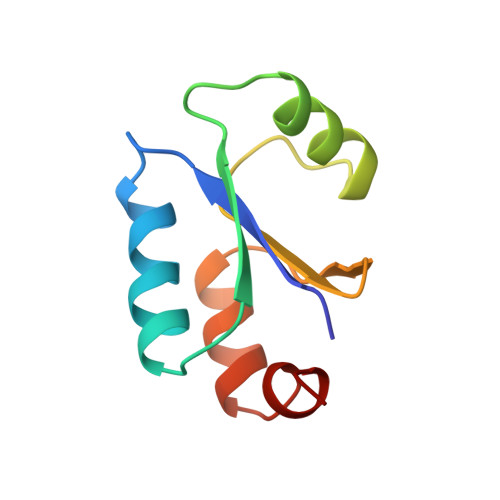
NMR structure of Escherichia coli glutaredoxin 3-glutathione mixed disulfide complex: implications for the enzymatic mechanism.

Nordstrand, K.slund, F.Holmgren, A.Otting, G.Berndt, K.D.

(1999) J Mol Biology 286: 541-552

  • DOI: https://doi.org/10.1006/jmbi.1998.2444
  • Primary Citation Related Structures: 
    3GRX

  • PubMed Abstract: 

    Glutaredoxins (Grxs) catalyze reversible oxidation/reduction of protein disulfide groups and glutathione-containing mixed disulfide groups via an active site Grx-glutathione mixed disulfide (Grx-SG) intermediate. The NMR solution structure of the Escherichia coli Grx3 mixed disulfide with glutathione (Grx3-SG) was determined using a C14S mutant which traps this intermediate in the redox reaction. The structure contains a thioredoxin fold, with a well-defined binding site for glutathione which involves two intermolecular backbone-backbone hydrogen bonds forming an antiparallel intermolecular beta-bridge between the protein and glutathione. The solution structure of E. coli Grx3-SG also suggests a binding site for a second glutathione in the reduction of the Grx3-SG intermediate, which is consistent with the specificity of reduction observed in Grxs. Molecular details of the structure in relation to the stability of the intermediate and the activity of Grx3 as a reductant of glutathione mixed disulfide groups are discussed. A comparison of glutathione binding in Grx3-SG and ligand binding in other members of the thioredoxin superfamily is presented, which illustrates the highly conserved intermolecular interactions in this protein family.


  • Organizational Affiliation
    • Department of Medical Biochemistry and Biophysics, Karolinska Institute, S-171 77, Stockholm, Sweden.

Macromolecule Content 

  • Total Structure Weight: 9.37 kDa 
  • Atom Count: 657 
  • Modeled Residue Count: 82 
  • Deposited Residue Count: 82 
  • Unique protein chains: 1

Macromolecules

Find similar proteins by:|  3D Structure
Entity ID: 1
MoleculeChains  Sequence LengthOrganismDetailsImage
GLUTAREDOXIN 382Escherichia coliMutation(s): 2 
UniProt
Find proteins for P0AC62 (Escherichia coli (strain K12))
Explore P0AC62 
Go to UniProtKB:  P0AC62
Entity Groups
Sequence Clusters30% Identity50% Identity70% Identity90% Identity95% Identity100% Identity
UniProt GroupP0AC62
Sequence Annotations
Expand
Reference Sequence

Small Molecules

Ligands 1 Unique
IDChains Name / Formula / InChI Key2D Diagram3D Interactions
GSH

Query on GSH


Download:Ideal Coordinates CCD File
B [auth A]Glutathione
C10 H17 N3 O6 S
RWSXRVCMGQZWBV-WDSKDSINSA-N

Biologically Interesting Molecules (External Reference) 

1 Unique
Entity ID: 2
IDChains NameType/Class2D Diagram3D Interactions
PRD_002593 (GSH)
Query on PRD_002593
B [auth A]GlutathionePeptide-like / Oxidation-reduction

Experimental Data & Validation

Experimental Data

  • Method: SOLUTION NMR
  • Conformers Calculated: 100 
  • Conformers Submitted: 20 
  • Selection Criteria: LEAST RESTRAINT VIOLATION 

Structure Validation

View Full Validation Report


Entry History 

Deposition Data

Revision History  (Full details and data files)

  • Version 1.0: 1999-03-30
    Type: Initial release
  • Version 1.1: 2008-03-25
    Changes: Version format compliance
  • Version 1.2: 2011-07-13
    Changes: Version format compliance
  • Version 1.3: 2012-01-18
    Changes: Non-polymer description
  • Version 1.4: 2018-03-14
    Changes: Database references, Other
  • Version 1.5: 2025-03-26
    Changes: Data collection, Database references, Derived calculations, Structure summary