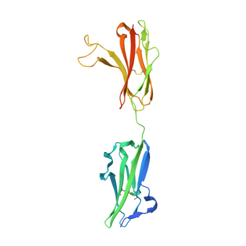
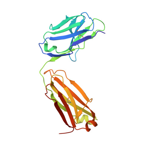
3GRW | pdb_00003grw

FGFR3 in complex with a Fab


Experimental Data Snapshot

  • Method: X-RAY DIFFRACTION
  • Resolution: 2.10 Å
  • R-Value Free: 
    0.228 (Depositor), 0.228 (DCC) 
  • R-Value Work: 
    0.189 (Depositor), 0.195 (DCC) 
  • R-Value Observed: 
    0.191 (Depositor) 

wwPDB Validation 3D Report Full Report

Validation slider image for 3GRW

Ligand Structure Quality Assessment 


This is version 1.3 of the entry. See complete history. 

Literature

Antibody-based targeting of FGFR3 in bladder carcinoma and t(4;14)-positive multiple myeloma in mice.

Qing, J., Du, X., Chen, Y., Chan, P., Li, H., Wu, P., Marsters, S., Stawicki, S., Tien, J., Totpal, K., Ross, S., Stinson, S., Dornan, D., French, D., Wang, Q.R., Stephan, J.P., Wu, Y., Wiesmann, C., Ashkenazi, A.

(2009) J Clin Invest 119: 1216-1229

  • DOI: https://doi.org/10.1172/JCI38017
  • Primary Citation Related Structures: 
    3GRW

  • PubMed Abstract: 

    Overexpression of FGF receptor 3 (FGFR3) is implicated in the development of t(4;14)-positive multiple myeloma. While FGFR3 is frequently overexpressed and/or activated through mutations in bladder cancer, the functional importance of FGFR3 and its potential as a specific therapeutic target in this disease have not been elucidated in vivo. Here we report that inducible knockdown of FGFR3 in human bladder carcinoma cells arrested cell-cycle progression in culture and markedly attenuated tumor progression in xenografted mice. Further, we developed a unique antibody (R3Mab) that inhibited not only WT FGFR3, but also various mutants of the receptor, including disulfide-linked cysteine mutants. Biochemical analysis and 2.1-A resolution crystallography revealed that R3Mab bound to a specific FGFR3 epitope that simultaneously blocked ligand binding, prevented receptor dimerization, and induced substantial conformational changes in the receptor. R3Mab exerted potent antitumor activity against bladder carcinoma and t(4;14)-positive multiple myeloma xenografts in mice by antagonizing FGFR3 signaling and eliciting antibody-dependent cell-mediated cytotoxicity (ADCC). These studies provide in vivo evidence demonstrating an oncogenic role of FGFR3 in bladder cancer and support antibody-based targeting of FGFR3 in hematologic and epithelial cancers driven by WT or mutant FGFR3.


  • Organizational Affiliation: 
    • Department of Molecular Oncology, Genentech Inc., South San Francisco, California, USA. jqing@gene.com

Macromolecule Content 

  • Total Structure Weight: 75.44 kDa 
  • Atom Count: 5,274 
  • Modeled Residue Count: 650 
  • Deposited Residue Count: 690 
  • Unique protein chains: 3

Macromolecules

Find similar proteins by:|  3D Structure
Entity ID: 1
MoleculeChains  Sequence LengthOrganismDetailsImage
Fibroblast growth factor receptor 3241Homo sapiensMutation(s): 0 
Gene Names: FGFR3
EC: 2.7.10.1
UniProt
Find proteins for Q8NI16 (Homo sapiens)
Explore Q8NI16 
Go to UniProtKB:  Q8NI16
Entity Groups
Sequence Clusters30% Identity50% Identity70% Identity90% Identity95% Identity100% Identity
UniProt GroupQ8NI16
Glycosylation
Glycosylation Sites: 1
Sequence Annotations
Expand
Reference Sequence
Find similar proteins by:|  3D Structure
Entity ID: 2
MoleculeChains  Sequence LengthOrganismDetailsImage
Fab light chainB [auth L]214Homo sapiensMutation(s): 0 
Entity Groups
Sequence Clusters30% Identity50% Identity70% Identity90% Identity95% Identity100% Identity
Sequence Annotations
Expand
Reference Sequence
Find similar proteins by:|  3D Structure
Entity ID: 3
MoleculeChains  Sequence LengthOrganismDetailsImage
Fab heavy chainC [auth H]235Homo sapiensMutation(s): 0 
Entity Groups
Sequence Clusters30% Identity50% Identity70% Identity90% Identity95% Identity100% Identity
Sequence Annotations
Expand
Reference Sequence

Small Molecules

Experimental Data & Validation

Experimental Data

  • Method: X-RAY DIFFRACTION
  • Resolution: 2.10 Å
  • R-Value Free:  0.228 (Depositor), 0.228 (DCC) 
  • R-Value Work:  0.189 (Depositor), 0.195 (DCC) 
  • R-Value Observed: 0.191 (Depositor) 
Space Group: P 21 21 21
Unit Cell:
Length ( Å )Angle ( ˚ )
a = 58.049α = 90
b = 99.329β = 90
c = 143.657γ = 90
Software Package:
Software NamePurpose
HKL-2000data collection
PHASERphasing
REFMACrefinement
DENZOdata reduction
SCALEPACKdata scaling

Structure Validation

View Full Validation Report


Ligand Structure Quality Assessment 


Entry History 

Deposition Data

  • Released Date: 2009-05-05 
  • Deposition Author(s): Wiesmann, C.

Revision History  (Full details and data files)

  • Version 1.0: 2009-05-05
    Type: Initial release
  • Version 1.1: 2011-07-13
    Changes: Advisory, Refinement description, Version format compliance
  • Version 1.2: 2020-07-29
    Type: Remediation
    Reason: Carbohydrate remediation
    Changes: Data collection, Database references, Derived calculations, Structure summary
  • Version 1.3: 2024-10-30
    Changes: Data collection, Database references, Structure summary