3GLJ | pdb_00003glj

A polymorph of carboxypeptidase B zymogen structure


Experimental Data Snapshot

  • Method: X-RAY DIFFRACTION
  • Resolution: 1.89 Å
  • R-Value Free: 
    0.198 (Depositor), 0.196 (DCC) 
  • R-Value Work: 
    0.166 (Depositor), 0.163 (DCC) 
  • R-Value Observed: 
    0.166 (Depositor) 

Starting Model: experimental
View more details

wwPDB Validation 3D Report Full Report

Validation slider image for 3GLJ

This is version 1.4 of the entry. See complete history

Literature

Analysis of a new crystal form of procarboxypeptidase B: further insights into the catalytic mechanism

Fernandez, D.Boix, E.Pallares, I.Aviles, F.X.Vendrell, J.

(2009) Biopolymers 

  • DOI: https://doi.org/10.1002/bip.21320
  • Primary Citation Related Structures: 
    3GLJ

  • PubMed Abstract: 

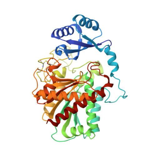
    A new triclinic crystal structure form of porcine pancreatic procarboxypeptidase B (PCPB) was obtained at higher resolution than the previously known tetragonal crystal structure. This new crystal polymorph has allowed for a corrected, accurate assignment of residues along the polypeptide chain based on the currently available gene sequence information and crystallographic data. The present structure shows unbound PCPB in a distinct molecular packing as compared to the previous benzamidine complexed form. Its catalytically important Tyr248 residue is oriented and hydrogen-bonded to solvent water molecules, and locates the furthest away from the catalytic zinc ion as compared to previous structures. A relatively long stretch of residues flanking Tyr248 and guarding the access to the catalytic zinc ion was found to be sequentially unique to the M14 family of peptidases. Predictions from a normal mode analysis indicated that this stretch of residues belongs to a rigid subdomain in the protein structure. The specific presence of a tyrosyl residue at the most exposed position in this region would allow for a delicate balance between extreme hydrophobicity and hydrophilicity, and affect substrate binding and the kinetic efficiency of the enzyme.


  • Organizational Affiliation
    • Departament de Bioquímica i Biologia Molecular, Facultat de Biociències, Universitat Autònoma de Barcelona, E-08193 Bellaterra, Spain.

Macromolecule Content 

  • Total Structure Weight: 46.41 kDa 
  • Atom Count: 3,511 
  • Modeled Residue Count: 395 
  • Deposited Residue Count: 401 
  • Unique protein chains: 1

Macromolecules

Find similar proteins by:|  3D Structure
Entity ID: 1
MoleculeChains  Sequence LengthOrganismDetailsImage
Carboxypeptidase B401Sus scrofaMutation(s): 0 
Gene Names: CPB1CPB
EC: 3.4.17.2
UniProt
Find proteins for P09955 (Sus scrofa)
Explore P09955 
Go to UniProtKB:  P09955
Entity Groups
Sequence Clusters30% Identity50% Identity70% Identity90% Identity95% Identity100% Identity
UniProt GroupP09955
Sequence Annotations
Expand
Reference Sequence

Experimental Data & Validation

Experimental Data

  • Method: X-RAY DIFFRACTION
  • Resolution: 1.89 Å
  • R-Value Free:  0.198 (Depositor), 0.196 (DCC) 
  • R-Value Work:  0.166 (Depositor), 0.163 (DCC) 
  • R-Value Observed: 0.166 (Depositor) 
Space Group: P 1
Unit Cell:
Length ( Å )Angle ( ˚ )
a = 49.59α = 109.94
b = 49.97β = 104.5
c = 50.63γ = 96.19
Software Package:
Software NamePurpose
XSCALEdata scaling
PHASERphasing
REFMACrefinement
PDB_EXTRACTdata extraction
MAR345dtbdata collection
XDSdata reduction

Structure Validation

View Full Validation Report


Entry History 

Deposition Data

Revision History  (Full details and data files)

  • Version 1.0: 2009-10-20
    Type: Initial release
  • Version 1.1: 2011-07-13
    Changes: Non-polymer description, Version format compliance
  • Version 1.2: 2017-02-08
    Changes: Other
  • Version 1.3: 2023-11-01
    Changes: Data collection, Database references, Derived calculations, Refinement description
  • Version 1.4: 2024-10-16
    Changes: Structure summary