3GJA | pdb_00003gja

CytC3


Experimental Data Snapshot

  • Method: X-RAY DIFFRACTION
  • Resolution: 2.20 Å
  • R-Value Free: 
    0.261 (Depositor), 0.260 (DCC) 
  • R-Value Work: 
    0.226 (Depositor), 0.225 (DCC) 

Starting Model: experimental
View more details

wwPDB Validation 3D Report Full Report

Validation slider image for 3GJA

This is version 1.3 of the entry. See complete history

Literature

Structural analysis of an open active site conformation of nonheme iron halogenase CytC3

Wong, C.Fujimori, D.G.Walsh, C.T.Drennan, C.L.

(2009) J Am Chem Soc 131: 4872-4879

  • DOI: https://doi.org/10.1021/ja8097355
  • Primary Citation Related Structures: 
    3GJA, 3GJB

  • PubMed Abstract: 

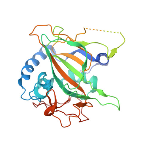
    CytC3, a member of the recently discovered class of nonheme Fe(II) and alpha-ketoglutarate (alphaKG)-dependent halogenases, catalyzes the double chlorination of L-2-aminobutyric acid (Aba) to produce a known Streptomyces antibiotic, gamma,gamma-dichloroaminobutyrate. Unlike the majority of the Fe(II)-alphaKG-dependent enzymes that catalyze hydroxylation reactions, halogenases catalyze a transfer of halides. To examine the important enzymatic features that discriminate between chlorination and hydroxylation, the crystal structures of CytC3 both with and without alphaKG/Fe(II) have been solved to 2.2 A resolution. These structures capture CytC3 in an open active site conformation, in which no chloride is bound to iron. Comparison of the open conformation of CytC3 with the closed conformation of another nonheme iron halogenase, SyrB2, suggests two important criteria for creating an enzyme-bound Fe-Cl catalyst: (1) the presence of a hydrogen-bonding network between the chloride and surrounding residues, and (2) the presence of a hydrophobic pocket in which the chloride resides.


  • Organizational Affiliation
    • Department of Chemistry, Massachusetts Institute of Technology, 77 Massachusetts Ave., Cambridge, Massachusetts 02139, USA.

Macromolecule Content 

  • Total Structure Weight: 73.21 kDa 
  • Atom Count: 4,546 
  • Modeled Residue Count: 538 
  • Deposited Residue Count: 638 
  • Unique protein chains: 1

Macromolecules

Find similar proteins by:|  3D Structure
Entity ID: 1
MoleculeChains  Sequence LengthOrganismDetailsImage
CytC3
A, B
319Streptomyces sp.Mutation(s): 0 
Gene Names: cytC3
UniProt
Find proteins for A0ACD6BAX6 (Streptomyces sp)
Explore A0ACD6BAX6 
Go to UniProtKB:  A0ACD6BAX6
Entity Groups
Sequence Clusters30% Identity50% Identity70% Identity90% Identity95% Identity100% Identity
UniProt GroupA0ACD6BAX6
Sequence Annotations
Expand
Reference Sequence

Experimental Data & Validation

Experimental Data

  • Method: X-RAY DIFFRACTION
  • Resolution: 2.20 Å
  • R-Value Free:  0.261 (Depositor), 0.260 (DCC) 
  • R-Value Work:  0.226 (Depositor), 0.225 (DCC) 
Space Group: P 43 21 2
Unit Cell:
Length ( Å )Angle ( ˚ )
a = 90.024α = 90
b = 90.024β = 90
c = 249.187γ = 90
Software Package:
Software NamePurpose
DENZOdata reduction
SCALEPACKdata scaling
PHASERphasing
CNSrefinement
PDB_EXTRACTdata extraction

Structure Validation

View Full Validation Report


Entry History 

Deposition Data

Revision History  (Full details and data files)

  • Version 1.0: 2009-04-14
    Type: Initial release
  • Version 1.1: 2011-07-13
    Changes: Derived calculations, Version format compliance
  • Version 1.2: 2017-11-01
    Changes: Refinement description
  • Version 1.3: 2023-09-06
    Changes: Data collection, Database references, Derived calculations, Refinement description