3GI1 | pdb_00003gi1

Crystal Structure of the laminin-binding protein Lbp of Streptococcus pyogenes


Experimental Data Snapshot

  • Method: X-RAY DIFFRACTION
  • Resolution: 2.45 Å
  • R-Value Free: 
    0.251 (Depositor), 0.243 (DCC) 
  • R-Value Work: 
    0.186 (Depositor), 0.180 (DCC) 
  • R-Value Observed: 
    0.190 (Depositor) 

Starting Models: experimental
View more details

wwPDB Validation 3D Report Full Report

Validation slider image for 3GI1

This is version 1.2 of the entry. See complete history

Literature

The laminin-binding protein Lbp from Streptococcus pyogenes is a zinc receptor

Linke, C.Caradoc-Davies, T.T.Young, P.G.Proft, T.Baker, E.N.

(2009) J Bacteriol 191: 5814-5823

  • DOI: https://doi.org/10.1128/JB.00485-09
  • Primary Citation Related Structures: 
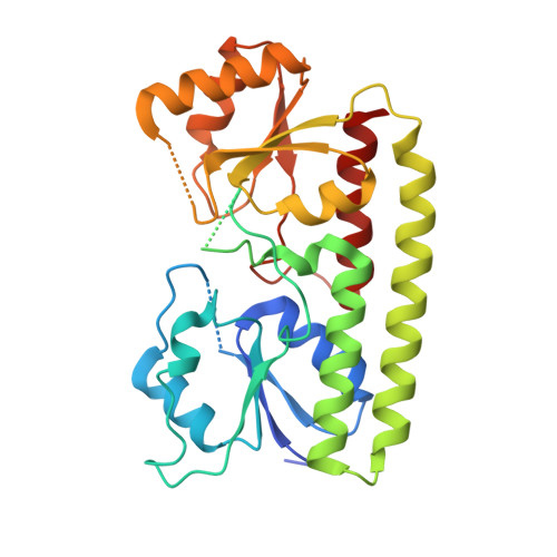
    3GI1

  • PubMed Abstract: 

    The common pathogen Streptococcus pyogenes colonizes the human skin and tonsils and can invade underlying tissues. This requires the adhesion of S. pyogenes to host surface receptors mediated through adhesins. The laminin-binding protein Lbp has been suggested as an adhesin, specific for the human extracellular matrix protein laminin. Sequence alignments, however, indicate a relationship between Lbp and a family of bacterial metal-binding receptors. To further analyze the role of Lbp in S. pyogenes and its potential role in pathogenicity, Lbp has been crystallized, and its structure has been solved at a resolution of 2.45 A (R = 0.186; R(free) = 0.251). Lbp has the typical metal-binding receptor fold, comprising two globular (beta/alpha)(4) domains connected by a helical backbone. The two domains enclose the metal-binding site, which contains a zinc ion. The interaction of Lbp with laminin was further investigated and shown to be specific in vitro. Localization studies with antibodies specific for Lbp show that the protein is attached to the membrane. The data suggest that Lbp is primarily a zinc-binding protein, and we suggest that its interaction with laminin in vivo may be mediated via zinc bound to laminin.


  • Organizational Affiliation
    • School of Biological Sciences, University of Auckland, Private Bag 92019, Auckland, New Zealand.

Macromolecule Content 

  • Total Structure Weight: 63.95 kDa 
  • Atom Count: 3,983 
  • Modeled Residue Count: 508 
  • Deposited Residue Count: 572 
  • Unique protein chains: 1

Macromolecules

Find similar proteins by:|  3D Structure
Entity ID: 1
MoleculeChains  Sequence LengthOrganismDetailsImage
Laminin-binding protein of group A streptococci
A, B
286Streptococcus pyogenesMutation(s): 0 
Gene Names: lmblmb/spy_2007
UniProt
Find proteins for Q99XV3 (Streptococcus pyogenes serotype M1)
Explore Q99XV3 
Go to UniProtKB:  Q99XV3
Entity Groups
Sequence Clusters30% Identity50% Identity70% Identity90% Identity95% Identity100% Identity
UniProt GroupQ99XV3
Sequence Annotations
Expand
Reference Sequence

Experimental Data & Validation

Experimental Data

  • Method: X-RAY DIFFRACTION
  • Resolution: 2.45 Å
  • R-Value Free:  0.251 (Depositor), 0.243 (DCC) 
  • R-Value Work:  0.186 (Depositor), 0.180 (DCC) 
  • R-Value Observed: 0.190 (Depositor) 
Space Group: P 1 21 1
Unit Cell:
Length ( Å )Angle ( ˚ )
a = 42.624α = 90
b = 92.163β = 106.27
c = 70.614γ = 90
Software Package:
Software NamePurpose
MOSFLMdata reduction
SCALAdata scaling
PHASERphasing
PHENIXrefinement
PDB_EXTRACTdata extraction

Structure Validation

View Full Validation Report


Entry History 

Deposition Data

Revision History  (Full details and data files)

  • Version 1.0: 2009-08-04
    Type: Initial release
  • Version 1.1: 2011-07-13
    Changes: Version format compliance
  • Version 1.2: 2023-11-01
    Changes: Data collection, Database references, Derived calculations, Refinement description