


Crystal structure of SAM-dependent methyltransferase (YP_325210.1) from ANABAENA VARIABILIS ATCC 29413 at 2.11 A resolution
Joint Center for Structural Genomics (JCSG)To be published.
Experimental Data Snapshot
Entity ID: 1 | |||||
|---|---|---|---|---|---|
| Molecule | Chains | Sequence Length | Organism | Details | Image |
| SAM-dependent methyltransferase | 245 | Trichormus variabilis ATCC 29413 | Mutation(s): 0 Gene Names: Ava_4718, YP_325210.1 | 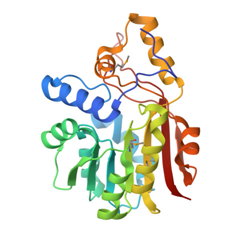 | |
UniProt | |||||
Entity Groups | |||||
| Sequence Clusters | 30% Identity50% Identity70% Identity90% Identity95% Identity100% Identity | ||||
| UniProt Group | Q3M3X1 | ||||
Sequence AnnotationsExpand | |||||
Reference Sequence | |||||
| Ligands 7 Unique | |||||
|---|---|---|---|---|---|
| ID | Chains | Name / Formula / InChI Key | 2D Diagram | 3D Interactions | |
| SAH Download:Ideal Coordinates CCD File | E [auth A] | S-ADENOSYL-L-HOMOCYSTEINE C14 H20 N6 O5 S ZJUKTBDSGOFHSH-WFMPWKQPSA-N |  | ||
| SO4 Download:Ideal Coordinates CCD File | G [auth A], H [auth A], I [auth A], J [auth A] | SULFATE ION O4 S QAOWNCQODCNURD-UHFFFAOYSA-L |  | ||
| IMD Download:Ideal Coordinates CCD File | F [auth A] | IMIDAZOLE C3 H5 N2 RAXXELZNTBOGNW-UHFFFAOYSA-O |  | ||
| EDO Download:Ideal Coordinates CCD File | K [auth A] L [auth A] M [auth A] N [auth A] O [auth A] | 1,2-ETHANEDIOL C2 H6 O2 LYCAIKOWRPUZTN-UHFFFAOYSA-N |  | ||
| CO Download:Ideal Coordinates CCD File | C [auth A] | COBALT (II) ION Co XLJKHNWPARRRJB-UHFFFAOYSA-N |  | ||
| CL Download:Ideal Coordinates CCD File | D [auth A] | CHLORIDE ION Cl VEXZGXHMUGYJMC-UHFFFAOYSA-M |  | ||
| UNL Download:Ideal Coordinates CCD File | B [auth A] | Unknown ligand VEXZGXHMUGYJMC-UHFFFAOYSA-M | |||
| Modified Residues 1 Unique | |||||
|---|---|---|---|---|---|
| ID | Chains | Type | Formula | 2D Diagram | Parent |
| MSE Query on MSE | A | L-PEPTIDE LINKING | C5 H11 N O2 Se |  | MET |
| Length ( Å ) | Angle ( ˚ ) |
|---|---|
| a = 112.52 | α = 90 |
| b = 112.52 | β = 90 |
| c = 112.56 | γ = 90 |
| Software Name | Purpose |
|---|---|
| REFMAC | refinement |
| PHENIX | refinement |
| SHELX | phasing |
| MolProbity | model building |
| XSCALE | data scaling |
| PDB_EXTRACT | data extraction |
| XDS | data reduction |
| SHELXD | phasing |
| autoSHARP | phasing |


