3GBM | pdb_00003gbm

Crystal Structure of Fab CR6261 in Complex with a H5N1 influenza virus hemagglutinin.


Experimental Data Snapshot

  • Method: X-RAY DIFFRACTION
  • Resolution: 2.70 Å
  • R-Value Free: 
    0.261 (Depositor), 0.272 (DCC) 
  • R-Value Work: 
    0.203 (Depositor) 
  • R-Value Observed: 
    0.206 (Depositor) 

Starting Models: experimental
View more details

wwPDB Validation 3D Report Full Report

Validation slider image for 3GBM

Ligand Structure Quality Assessment 


This is version 2.2 of the entry. See complete history

Literature

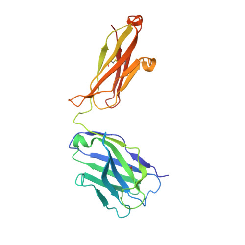
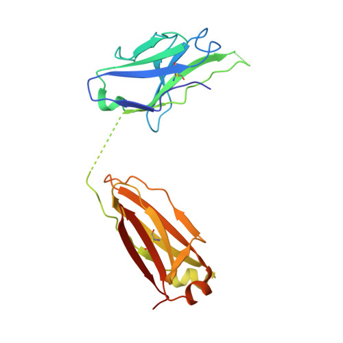
Antibody recognition of a highly conserved influenza virus epitope.

Ekiert, D.C.Bhabha, G.Elsliger, M.A.Friesen, R.H.Jongeneelen, M.Throsby, M.Goudsmit, J.Wilson, I.A.

(2009) Science 324: 246-251

  • DOI: https://doi.org/10.1126/science.1171491
  • Primary Citation Related Structures: 
    3GBM, 3GBN

  • PubMed Abstract: 

    Influenza virus presents an important and persistent threat to public health worldwide, and current vaccines provide immunity to viral isolates similar to the vaccine strain. High-affinity antibodies against a conserved epitope could provide immunity to the diverse influenza subtypes and protection against future pandemic viruses. Cocrystal structures were determined at 2.2 and 2.7 angstrom resolutions for broadly neutralizing human antibody CR6261 Fab in complexes with the major surface antigen (hemagglutinin, HA) from viruses responsible for the 1918 H1N1 influenza pandemic and a recent lethal case of H5N1 avian influenza. In contrast to other structurally characterized influenza antibodies, CR6261 recognizes a highly conserved helical region in the membrane-proximal stem of HA1 and HA2. The antibody neutralizes the virus by blocking conformational rearrangements associated with membrane fusion. The CR6261 epitope identified here should accelerate the design and implementation of improved vaccines that can elicit CR6261-like antibodies, as well as antibody-based therapies for the treatment of influenza.


  • Organizational Affiliation
    • Department of Molecular Biology, Scripps Research Institute, 10550 North Torrey Pines Road, La Jolla, CA 92037, USA.

Macromolecule Content 

  • Total Structure Weight: 213.73 kDa 
  • Atom Count: 14,189 
  • Modeled Residue Count: 1,829 
  • Deposited Residue Count: 1,916 
  • Unique protein chains: 4

Macromolecules

Find similar proteins by:|  3D Structure
Entity ID: 1
MoleculeChains  Sequence LengthOrganismDetailsImage
Hemagglutinin
A, C
334Influenza A virus (A/Viet Nam/1203/2004(H5N1))Mutation(s): 0 
Gene Names: HAHemagglutinin (HA)
UniProt
Find proteins for Q6DQ33 (Influenza A virus (strain A/Vietnam/1203/2004 H5N1))
Explore Q6DQ33 
Go to UniProtKB:  Q6DQ33
Entity Groups
Sequence Clusters30% Identity50% Identity70% Identity90% Identity95% Identity100% Identity
UniProt GroupQ6DQ33
Glycosylation
Glycosylation Sites: 3
Sequence Annotations
Expand
Reference Sequence
Find similar proteins by:|  3D Structure
Entity ID: 2
MoleculeChains  Sequence LengthOrganismDetailsImage
Hemagglutinin
B, D
177Influenza A virus (A/Viet Nam/1203/2004(H5N1))Mutation(s): 0 
Gene Names: HAHemagglutinin (HA)
UniProt
Find proteins for Q6DQ33 (Influenza A virus (strain A/Vietnam/1203/2004 H5N1))
Explore Q6DQ33 
Go to UniProtKB:  Q6DQ33
Entity Groups
Sequence Clusters30% Identity50% Identity70% Identity90% Identity95% Identity100% Identity
UniProt GroupQ6DQ33
Glycosylation
Glycosylation Sites: 1
Sequence Annotations
Expand
Reference Sequence
Find similar proteins by:|  3D Structure
Entity ID: 3
MoleculeChains  Sequence LengthOrganismDetailsImage
antibody (Fab)E [auth H],
G [auth I]
226Homo sapiensMutation(s): 0 
Find similar proteins by:|  3D Structure
Entity ID: 4
MoleculeChains  Sequence LengthOrganismDetailsImage
antibody (Fab)F [auth L],
H [auth M]
221Homo sapiensMutation(s): 0 

Oligosaccharides

Help  
Entity ID: 5
MoleculeChains Length2D Diagram GlycosylationD Interactions
2-acetamido-2-deoxy-beta-D-glucopyranose-(1-4)-2-acetamido-2-deoxy-beta-D-glucopyranoseI [auth E],
J [auth F]
2N-Glycosylation
Glycosylation Resources
GlyTouCan: G42666HT
GlyCosmos: G42666HT
GlyGen: G42666HT

Small Molecules

Ligands 4 Unique
IDChains Name / Formula / InChI Key2D Diagram3D Interactions
NAG

Query on NAG


Download:Ideal Coordinates CCD File
K [auth A],
M [auth A],
O [auth B],
R [auth C],
T [auth D]
2-acetamido-2-deoxy-beta-D-glucopyranose
C8 H15 N O6
OVRNDRQMDRJTHS-FMDGEEDCSA-N
BMA

Query on BMA


Download:Ideal Coordinates CCD File
L [auth A],
Q [auth C]
beta-D-mannopyranose
C6 H12 O6
WQZGKKKJIJFFOK-RWOPYEJCSA-N
GOL

Query on GOL


Download:Ideal Coordinates CCD File
P [auth B]GLYCEROL
C3 H8 O3
PEDCQBHIVMGVHV-UHFFFAOYSA-N
EDO

Query on EDO


Download:Ideal Coordinates CCD File
N [auth A],
S [auth C]
1,2-ETHANEDIOL
C2 H6 O2
LYCAIKOWRPUZTN-UHFFFAOYSA-N

Experimental Data & Validation

Experimental Data

  • Method: X-RAY DIFFRACTION
  • Resolution: 2.70 Å
  • R-Value Free:  0.261 (Depositor), 0.272 (DCC) 
  • R-Value Work:  0.203 (Depositor) 
  • R-Value Observed: 0.206 (Depositor) 
Space Group: P 21 3
Unit Cell:
Length ( Å )Angle ( ˚ )
a = 202.761α = 90
b = 202.761β = 90
c = 202.761γ = 90
Software Package:
Software NamePurpose
Blu-Icedata collection
PHASERphasing
REFMACrefinement
HKL-2000data reduction
SCALEPACKdata scaling

Structure Validation

View Full Validation Report


Ligand Structure Quality Assessment 


Entry History 

Deposition Data

Revision History  (Full details and data files)

  • Version 1.0: 2009-03-10
    Type: Initial release
  • Version 1.1: 2011-07-13
    Changes: Advisory, Version format compliance
  • Version 2.0: 2020-07-29
    Type: Remediation
    Reason: Carbohydrate remediation
    Changes: Advisory, Atomic model, Data collection, Derived calculations, Structure summary
  • Version 2.1: 2023-09-06
    Changes: Data collection, Database references, Derived calculations, Refinement description, Structure summary
  • Version 2.2: 2024-10-16
    Changes: Structure summary