


Crystal structure of the motor domain of kinesin KIF13B bound with ADP
Tong, Y., Shen, L., Shen, Y., Tempel, W., Arrowsmith, C.H., Edwards, A.M., Bountra, C., Weigelt, J., Bochkarev, A., Park, H.To be published.
Experimental Data Snapshot
Starting Model: experimental
View more details
Entity ID: 1 | |||||
|---|---|---|---|---|---|
| Molecule | Chains | Sequence Length | Organism | Details | Image |
| KIF13B protein | 354 | Homo sapiens | Mutation(s): 0 Gene Names: KIF13B | 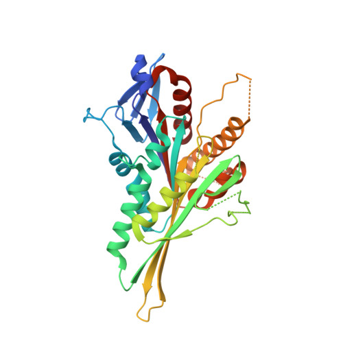 | |
UniProt & NIH Common Fund Data Resources | |||||
PHAROS: Q9NQT8 GTEx: ENSG00000197892 | |||||
Entity Groups | |||||
| Sequence Clusters | 30% Identity50% Identity70% Identity90% Identity95% Identity100% Identity | ||||
| UniProt Group | Q9NQT8 | ||||
Sequence AnnotationsExpand | |||||
Reference Sequence | |||||
| Ligands 3 Unique | |||||
|---|---|---|---|---|---|
| ID | Chains | Name / Formula / InChI Key | 2D Diagram | 3D Interactions | |
| ADP Download:Ideal Coordinates CCD File | D [auth A], H [auth B], L [auth C] | ADENOSINE-5'-DIPHOSPHATE C10 H15 N5 O10 P2 XTWYTFMLZFPYCI-KQYNXXCUSA-N |  | ||
| MG Download:Ideal Coordinates CCD File | E [auth A], I [auth B], M [auth C] | MAGNESIUM ION Mg JLVVSXFLKOJNIY-UHFFFAOYSA-N |  | ||
| UNX Download:Ideal Coordinates CCD File | F [auth A], G [auth A], J [auth B], K [auth B], N [auth C] | UNKNOWN ATOM OR ION X |  | ||
| Length ( Å ) | Angle ( ˚ ) |
|---|---|
| a = 74.229 | α = 90 |
| b = 86.52 | β = 106.8 |
| c = 94.948 | γ = 90 |
| Software Name | Purpose |
|---|---|
| DENZO | data reduction |
| SCALEPACK | data scaling |
| PHASER | phasing |
| PHENIX | refinement |
| PDB_EXTRACT | data extraction |
| ADSC | data collection |
| HKL-3000 | data reduction |
| HKL-3000 | data scaling |


