3G2T

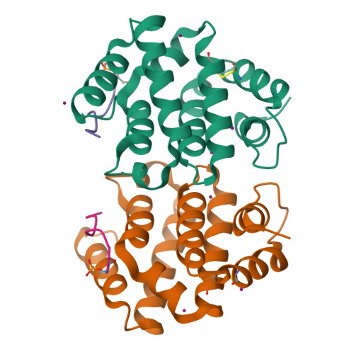
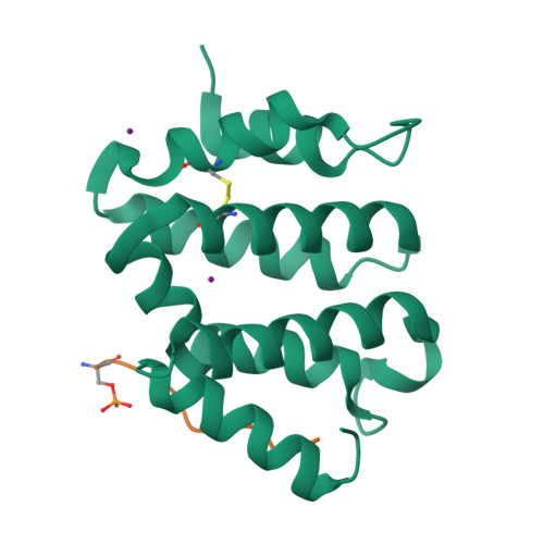
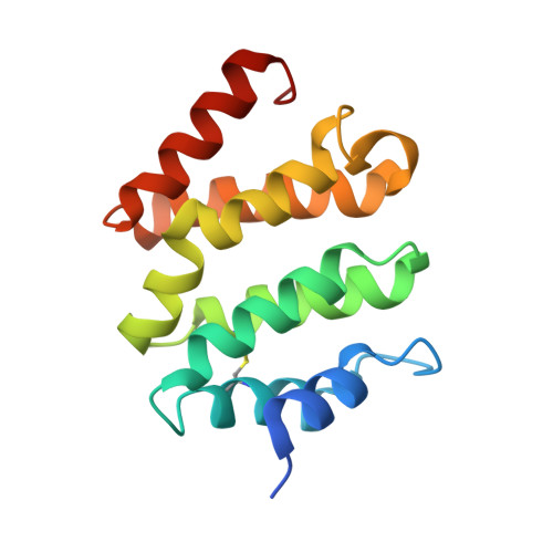
VHS Domain of human GGA1 complexed with SorLA C-terminal Phosphopeptide


Experimental Data Snapshot

  • Method: X-RAY DIFFRACTION
  • Resolution: 2.00 Å
  • R-Value Free: 0.233 
  • R-Value Work: 0.192 
  • R-Value Observed: 0.194 

Starting Model: experimental
View more details

wwPDB Validation   3D Report Full Report


This is version 1.4 of the entry. See complete history


Literature

GGA autoinhibition revisited

Cramer, J.F.Gustafsen, C.Behrens, M.A.Oliveira, C.L.P.Pedersen, J.S.Madsen, P.Petersen, C.M.Thirup, S.S.

(2010) Traffic 11: 259-273

  • DOI: https://doi.org/10.1111/j.1600-0854.2009.01017.x
  • Primary Citation of Related Structures:  
    3G2S, 3G2T, 3G2U, 3G2V, 3G2W

  • PubMed Abstract: 

    The cytosolic adaptors GGA1-3 mediate sorting of transmembrane proteins displaying a C-terminal acidic dileucine motif (DXXLL) in their cytosolic domain. GGA1 and GGA3 contain similar but intrinsic motifs that are believed to serve as autoinhibitory sites activated by the phosphorylation of a serine positioned three residues upstream of the DXXLL motif. In the present study, we have subjected the widely acknowledged concept of GGA1 autoinhibition to a thorough structural and functional examination. We find that (i) the intrinsic motif of GGA1 is inactive, (ii) only C-terminal DXXLL motifs constitute active GGA binding sites, (iii) while aspartates and phosphorylated serines one or two positions upstream of the DXXLL motif increase GGA1 binding, phosphoserines further upstream have little or no influence and (iv) phosphorylation of GGA1 does not affect its conformation or binding to Sortilin and SorLA. Taken together, our findings seem to refute the functional significance of GGA autoinhibition in particular and of intrinsic GGA binding motifs in general.


  • Organizational Affiliation

    MIND Centre, Department of Molecular Biology, Aarhus University, Gustav Wieds Vej 10 C, DK-8000 Aarhus C, Denmark.


Macromolecules
Find similar proteins by:  (by identity cutoff)  |  3D Structure
Entity ID: 1
MoleculeChains Sequence LengthOrganismDetailsImage
ADP-ribosylation factor-binding protein GGA1
A, B
149Homo sapiensMutation(s): 0 
UniProt & NIH Common Fund Data Resources
Find proteins for Q9UJY5 (Homo sapiens)
Explore Q9UJY5 
Go to UniProtKB:  Q9UJY5
PHAROS:  Q9UJY5
GTEx:  ENSG00000100083 
Entity Groups  
Sequence Clusters30% Identity50% Identity70% Identity90% Identity95% Identity100% Identity
UniProt GroupQ9UJY5
Sequence Annotations
Expand
  • Reference Sequence

Find similar proteins by:  Sequence   |   3D Structure  

Entity ID: 2
MoleculeChains Sequence LengthOrganismDetailsImage
Phosphorylated C-terminal fragment of Sortilin-related receptor
C, D
13N/AMutation(s): 0 
UniProt & NIH Common Fund Data Resources
Find proteins for Q92673 (Homo sapiens)
Explore Q92673 
Go to UniProtKB:  Q92673
PHAROS:  Q92673
GTEx:  ENSG00000137642 
Entity Groups  
Sequence Clusters30% Identity50% Identity70% Identity90% Identity95% Identity100% Identity
UniProt GroupQ92673
Sequence Annotations
Expand
  • Reference Sequence
Small Molecules
Modified Residues  1 Unique
IDChains TypeFormula2D DiagramParent
SEP
Query on SEP
C, D
L-PEPTIDE LINKINGC3 H8 N O6 PSER
Experimental Data & Validation

Experimental Data

  • Method: X-RAY DIFFRACTION
  • Resolution: 2.00 Å
  • R-Value Free: 0.233 
  • R-Value Work: 0.192 
  • R-Value Observed: 0.194 
  • Space Group: P 21 21 21
Unit Cell:
Length ( Å )Angle ( ˚ )
a = 55.8α = 90
b = 68.3β = 90
c = 100.6γ = 90
Software Package:
Software NamePurpose
PHENIXrefinement
PDB_EXTRACTdata extraction
XDSdata scaling
XDSdata reduction
XSCALEdata scaling
PHASERphasing

Structure Validation

View Full Validation Report


Entry History 

Deposition Data

Revision History  (Full details and data files)

  • Version 1.0: 2009-12-15
    Type: Initial release
  • Version 1.1: 2011-07-13
    Changes: Version format compliance
  • Version 1.2: 2014-02-05
    Changes: Database references
  • Version 1.3: 2023-11-01
    Changes: Data collection, Database references, Derived calculations, Refinement description
  • Version 1.4: 2024-11-13
    Changes: Structure summary