3FY4 | pdb_00003fy4

(6-4) Photolyase Crystal Structure


Experimental Data Snapshot

  • Method: X-RAY DIFFRACTION
  • Resolution: 2.70 Å
  • R-Value Free: 
    0.237 (Depositor), 0.229 (DCC) 
  • R-Value Work: 
    0.202 (Depositor), 0.195 (DCC) 

wwPDB Validation 3D Report Full Report

Validation slider image for 3FY4

Ligand Structure Quality Assessment 


This is version 1.2 of the entry. See complete history

Literature

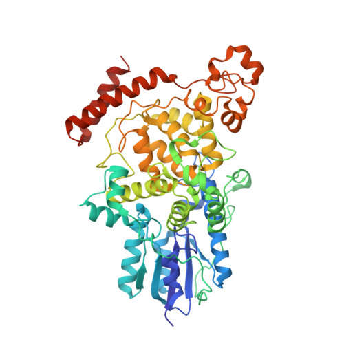
Functional motifs in the (6-4) photolyase crystal structure make a comparative framework for DNA repair photolyases and clock cryptochromes.

Hitomi, K.DiTacchio, L.Arvai, A.S.Yamamoto, J.Kim, S.T.Todo, T.Tainer, J.A.Iwai, S.Panda, S.Getzoff, E.D.

(2009) Proc Natl Acad Sci U S A 106: 6962-6967

  • DOI: https://doi.org/10.1073/pnas.0809180106
  • Primary Citation Related Structures: 
    3FY4

  • PubMed Abstract: 

    Homologous flavoproteins from the photolyase (PHR)/cryptochrome (CRY) family use the FAD cofactor in PHRs to catalyze DNA repair and in CRYs to tune the circadian clock and control development. To help address how PHR/CRY members achieve these diverse functions, we determined the crystallographic structure of Arabidopsis thaliana (6-4) PHR (UVR3), which is strikingly (>65%) similar in sequence to human circadian clock CRYs. The structure reveals a substrate-binding cavity specific for the UV-induced DNA lesion, (6-4) photoproduct, and cofactor binding sites different from those of bacterial PHRs and consistent with distinct mechanisms for activities and regulation. Mutational analyses were combined with this prototypic structure for the (6-4) PHR/clock CRY cluster to identify structural and functional motifs: phosphate-binding and Pro-Lys-Leu protrusion motifs constricting access to the substrate-binding cavity above FAD, sulfur loop near the external end of the Trp electron-transfer pathway, and previously undefined C-terminal helix. Our results provide a detailed, unified framework for investigations of (6-4) PHRs and the mammalian CRYs. Conservation of key residues and motifs controlling FAD access and activities suggests that regulation of FAD redox properties and radical stability is essential not only for (6-4) photoproduct DNA repair, but also for circadian clock-regulating CRY functions. The structural and functional results reported here elucidate archetypal relationships within this flavoprotein family and suggest how PHRs and CRYs use local residue and cofactor tuning, rather than larger structural modifications, to achieve their diverse functions encompassing DNA repair, plant growth and development, and circadian clock regulation.


  • Organizational Affiliation
    • Graduate School of Engineering Science, Osaka University, Toyonaka, Osaka 560-8531, Japan.

Macromolecule Content 

  • Total Structure Weight: 189.04 kDa 
  • Atom Count: 13,491 
  • Modeled Residue Count: 1,563 
  • Deposited Residue Count: 1,611 
  • Unique protein chains: 1

Macromolecules

Find similar proteins by:|  3D Structure
Entity ID: 1
MoleculeChains  Sequence LengthOrganismDetailsImage
6-4 photolyase
A, B, C
537Arabidopsis thalianaMutation(s): 0 
Gene Names: 6-4 PHOTOLYASEUVR3
EC: 4.1.99.13
UniProt
Find proteins for O48652 (Arabidopsis thaliana)
Explore O48652 
Go to UniProtKB:  O48652
Entity Groups
Sequence Clusters30% Identity50% Identity70% Identity90% Identity95% Identity100% Identity
UniProt GroupO48652
Sequence Annotations
Expand
Reference Sequence

Small Molecules

Ligands 4 Unique
IDChains Name / Formula / InChI Key2D Diagram3D Interactions
FAD

Query on FAD


Download:Ideal Coordinates CCD File
I [auth A],
Q [auth B],
V [auth C]
FLAVIN-ADENINE DINUCLEOTIDE
C27 H33 N9 O15 P2
VWWQXMAJTJZDQX-UYBVJOGSSA-N
MES

Query on MES


Download:Ideal Coordinates CCD File
G [auth A],
O [auth B],
T [auth C]
2-(N-MORPHOLINO)-ETHANESULFONIC ACID
C6 H13 N O4 S
SXGZJKUKBWWHRA-UHFFFAOYSA-N
PO4

Query on PO4


Download:Ideal Coordinates CCD File
H [auth A],
P [auth B],
U [auth C]
PHOSPHATE ION
O4 P
NBIIXXVUZAFLBC-UHFFFAOYSA-K
IMD

Query on IMD


Download:Ideal Coordinates CCD File
D [auth A]
E [auth A]
F [auth A]
J [auth B]
K [auth B]
D [auth A],
E [auth A],
F [auth A],
J [auth B],
K [auth B],
L [auth B],
M [auth B],
N [auth B],
R [auth C],
S [auth C]
IMIDAZOLE
C3 H5 N2
RAXXELZNTBOGNW-UHFFFAOYSA-O

Experimental Data & Validation

Experimental Data

  • Method: X-RAY DIFFRACTION
  • Resolution: 2.70 Å
  • R-Value Free:  0.237 (Depositor), 0.229 (DCC) 
  • R-Value Work:  0.202 (Depositor), 0.195 (DCC) 
Space Group: P 21 21 21
Unit Cell:
Length ( Å )Angle ( ˚ )
a = 112.447α = 90
b = 139.033β = 90
c = 143.118γ = 90
Software Package:
Software NamePurpose
CNSrefinement
PDB_EXTRACTdata extraction
HKL-2000data collection
HKL-2000data reduction
SCALEPACKdata scaling
AMoREphasing

Structure Validation

View Full Validation Report


Ligand Structure Quality Assessment 


Entry History 

Deposition Data

Revision History  (Full details and data files)

  • Version 1.0: 2009-04-28
    Type: Initial release
  • Version 1.1: 2011-07-13
    Changes: Version format compliance
  • Version 1.2: 2024-02-21
    Changes: Data collection, Database references, Derived calculations