3FW0 | pdb_00003fw0

Structure of Peptidyl-alpha-hydroxyglycine alpha-Amidating Lyase (PAL) bound to alpha-hydroxyhippuric acid (non-peptidic substrate)


Experimental Data Snapshot

  • Method: X-RAY DIFFRACTION
  • Resolution: 2.52 Å
  • R-Value Free: 
    0.260 (Depositor), 0.303 (DCC) 
  • R-Value Work: 
    0.207 (Depositor), 0.258 (DCC) 
  • R-Value Observed: 
    0.209 (Depositor) 

Starting Model: experimental
View more details

wwPDB Validation 3D Report Full Report

Validation slider image for 3FW0

Ligand Structure Quality Assessment 


This is version 1.3 of the entry. See complete history

Literature

Amidation of bioactive peptides: the structure of the lyase domain of the amidating enzyme.

Chufan, E.E.De, M.Eipper, B.A.Mains, R.E.Amzel, L.M.

(2009) Structure 17: 965-973

  • DOI: https://doi.org/10.1016/j.str.2009.05.008
  • Primary Citation Related Structures: 
    3FVZ, 3FW0

  • PubMed Abstract: 

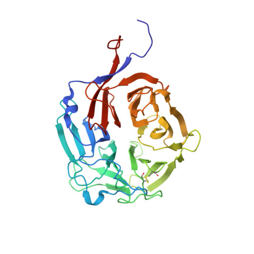
    Many neuropeptides and peptide hormones require amidation of their carboxy terminal for full biological activity. The enzyme peptidyl-alpha-hydroxyglycine alpha-amidating lyase (PAL; EC 4.3.2.5) catalyzes the second and last step of this reaction, N-dealkylation of the peptidyl-alpha-hydroxyglycine to generate the alpha-amidated peptide and glyoxylate. Here we report the X-ray crystal structure of the PAL catalytic core (PALcc) alone and in complex with the nonpeptidic substrate alpha-hydroxyhippuric acid. The structures show that PAL folds as a six-bladed beta-propeller. The active site is formed by a Zn(II) ion coordinated by three histidine residues; the substrate binds to this site with its alpha-hydroxyl group coordinated to the Zn(II) ion. The structures also reveal a tyrosine residue (Tyr(654)) at the active site as the catalytic base for hydroxyl deprotonation, an unusual role for tyrosine. A reaction mechanism is proposed based on this structural data and validated by biochemical analysis of site-directed PALcc mutants.


  • Organizational Affiliation
    • Department of Biophysics and Biophysical Chemistry, Johns Hopkins School of Medicine, Johns Hopkins University, Baltimore, MD 21205, USA.

Macromolecule Content 

  • Total Structure Weight: 37.55 kDa 
  • Atom Count: 2,754 
  • Modeled Residue Count: 329 
  • Deposited Residue Count: 329 
  • Unique protein chains: 1

Macromolecules

Find similar proteins by:|  3D Structure
Entity ID: 1
MoleculeChains  Sequence LengthOrganismDetailsImage
Peptidyl-glycine alpha-amidating monooxygenase329Rattus norvegicusMutation(s): 0 
Gene Names: Pam
EC: 4.3.2.5 (PDB Primary Data), 1.14.17.3 (UniProt)
UniProt
Find proteins for P14925 (Rattus norvegicus)
Explore P14925 
Go to UniProtKB:  P14925
Entity Groups
Sequence Clusters30% Identity50% Identity70% Identity90% Identity95% Identity100% Identity
UniProt GroupP14925
Sequence Annotations
Expand
Reference Sequence

Experimental Data & Validation

Experimental Data

  • Method: X-RAY DIFFRACTION
  • Resolution: 2.52 Å
  • R-Value Free:  0.260 (Depositor), 0.303 (DCC) 
  • R-Value Work:  0.207 (Depositor), 0.258 (DCC) 
  • R-Value Observed: 0.209 (Depositor) 
Space Group: P 21 21 21
Unit Cell:
Length ( Å )Angle ( ˚ )
a = 51.93α = 90
b = 75.08β = 90
c = 97.026γ = 90
Software Package:
Software NamePurpose
AMoREphasing
REFMACrefinement
HKL-2000data reduction
HKL-2000data scaling

Structure Validation

View Full Validation Report


Ligand Structure Quality Assessment 


Entry History 

Deposition Data

Revision History  (Full details and data files)

  • Version 1.0: 2009-09-01
    Type: Initial release
  • Version 1.1: 2011-07-13
    Changes: Version format compliance
  • Version 1.2: 2024-04-03
    Changes: Data collection, Database references, Derived calculations, Refinement description
  • Version 1.3: 2024-11-06
    Changes: Structure summary