3FRY | pdb_00003fry

Crystal structure of the CopA C-terminal metal binding domain


Experimental Data Snapshot

  • Method: X-RAY DIFFRACTION
  • Resolution: 2.00 Å
  • R-Value Free: 
    0.238 (Depositor) 
  • R-Value Work: 
    0.222 (Depositor), 0.234 (DCC) 
  • R-Value Observed: 
    0.222 (Depositor) 

Starting Model: experimental
View more details

wwPDB Validation 3D Report Full Report

Validation slider image for 3FRY

This is version 1.4 of the entry. See complete history

Literature

Structure and interactions of the C-terminal metal binding domain of Archaeoglobus fulgidus CopA.

Agarwal, S.Hong, D.Desai, N.K.Sazinsky, M.H.Arguello, J.M.Rosenzweig, A.C.

(2010) Proteins 78: 2450-2458

  • DOI: https://doi.org/10.1002/prot.22753
  • Primary Citation Related Structures: 
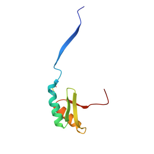
    3FRY

  • PubMed Abstract: 

    The Cu(+)-ATPase CopA from Archaeoglobus fulgidus belongs to the P(1B) family of the P-type ATPases. These integral membrane proteins couple the energy of ATP hydrolysis to heavy metal ion translocation across membranes. A defining feature of P(1B-1)-type ATPases is the presence of soluble metal binding domains at the N-terminus (N-MBDs). The N-MBDs exhibit a conserved ferredoxin-like fold, similar to that of soluble copper chaperones, and bind metal ions via a conserved CXXC motif. The N-MBDs enable Cu(+) regulation of turnover rates apparently through Cu-sensitive interactions with catalytic domains. A. fulgidus CopA is unusual in that it contains both an N-terminal MBD and a C-terminal MBD (C-MBD). The functional role of the unique C-MBD has not been established. Here, we report the crystal structure of the apo, oxidized C-MBD to 2.0 A resolution. In the structure, two C-MBD monomers form a domain-swapped dimer, which has not been observed previously for similar domains. In addition, the interaction of the C-MBD with the other cytoplasmic domains of CopA, the ATP binding domain (ATPBD) and actuator domain (A-domain), has been investigated. Interestingly, the C-MBD interacts specifically with both of these domains, independent of the presence of Cu(+) or nucleotides. These data reinforce the uniqueness of the C-MBD and suggest a distinct structural role for the C-MBD in CopA transport.


  • Organizational Affiliation
    • Department of Biochemistry, Molecular Biology, and Cell Biology, Northwestern University, Evanston, Illinois 60208, USA.

Macromolecule Content 

  • Total Structure Weight: 15.84 kDa 
  • Atom Count: 1,149 
  • Modeled Residue Count: 145 
  • Deposited Residue Count: 146 
  • Unique protein chains: 1

Macromolecules

Find similar proteins by:|  3D Structure
Entity ID: 1
MoleculeChains  Sequence LengthOrganismDetailsImage
Probable copper-exporting P-type ATPase A
A, B
73Archaeoglobus fulgidusMutation(s): 0 
Gene Names: AF_0473copApacS
EC: 3.6.3 (PDB Primary Data), 7.2.2.8 (UniProt)
UniProt
Find proteins for O29777 (Archaeoglobus fulgidus (strain ATCC 49558 / DSM 4304 / JCM 9628 / NBRC 100126 / VC-16))
Explore O29777 
Go to UniProtKB:  O29777
Entity Groups
Sequence Clusters30% Identity50% Identity70% Identity90% Identity95% Identity100% Identity
UniProt GroupO29777
Sequence Annotations
Expand
Reference Sequence

Small Molecules

Ligands 1 Unique
IDChains Name / Formula / InChI Key2D Diagram3D Interactions
CIT

Query on CIT


Download:Ideal Coordinates CCD File
C [auth A]CITRIC ACID
C6 H8 O7
KRKNYBCHXYNGOX-UHFFFAOYSA-N

Experimental Data & Validation

Experimental Data

  • Method: X-RAY DIFFRACTION
  • Resolution: 2.00 Å
  • R-Value Free:  0.238 (Depositor) 
  • R-Value Work:  0.222 (Depositor), 0.234 (DCC) 
  • R-Value Observed: 0.222 (Depositor) 
Space Group: P 1 2 1
Unit Cell:
Length ( Å )Angle ( ˚ )
a = 40.131α = 90
b = 35.989β = 104.89
c = 51.262γ = 90
Software Package:
Software NamePurpose
CNSrefinement
HKL-2000data reduction
HKL-2000data scaling

Structure Validation

View Full Validation Report


Entry History 

Deposition Data

Revision History  (Full details and data files)

  • Version 1.0: 2010-01-12
    Type: Initial release
  • Version 1.1: 2011-07-13
    Changes: Version format compliance
  • Version 1.2: 2011-11-16
    Changes: Atomic model
  • Version 1.3: 2023-09-06
    Changes: Data collection, Database references, Refinement description
  • Version 1.4: 2024-10-16
    Changes: Structure summary