3FO2 | pdb_00003fo2

Crystal structure of hapten complex of catalytic elimination antibody 13G5 (Glu(L39)Gln mutant)


Experimental Data Snapshot

  • Method: X-RAY DIFFRACTION
  • Resolution: 2.18 Å
  • R-Value Free: 
    0.256 (Depositor), 0.264 (DCC) 
  • R-Value Work: 
    0.201 (Depositor), 0.212 (DCC) 
  • R-Value Observed: 
    0.204 (Depositor) 

Starting Model: experimental
View more details

wwPDB Validation 3D Report Full Report

Validation slider image for 3FO2

Ligand Structure Quality Assessment 


This is version 1.5 of the entry. See complete history

Literature

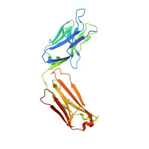
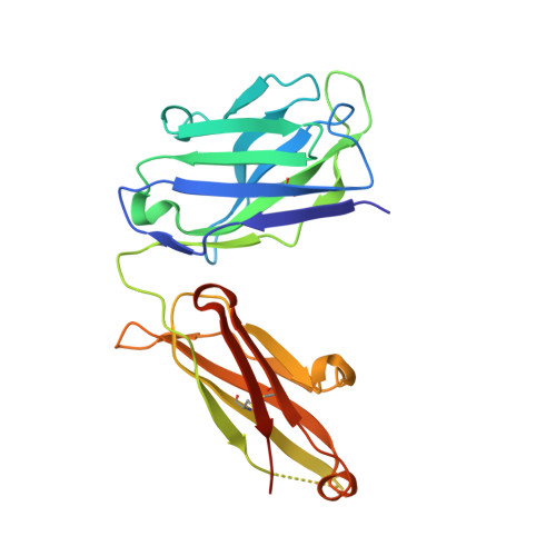
An aspartate and a water molecule mediate efficient acid-base catalysis in a tailored antibody pocket.

Debler, E.W.Muller, R.Hilvert, D.Wilson, I.A.

(2009) Proc Natl Acad Sci U S A 106: 18539-18544

  • DOI: https://doi.org/10.1073/pnas.0902700106
  • Primary Citation Related Structures: 
    3FO0, 3FO1, 3FO2

  • PubMed Abstract: 

    Design of catalysts featuring multiple functional groups is a desirable, yet formidable goal. Antibody 13G5, which accelerates the cleavage of unactivated benzisoxazoles, is one of few artificial enzymes that harness an acid and a base to achieve efficient proton transfer. X-ray structures of the Fab-hapten complexes of wild-type 13G5 and active-site variants now afford detailed insights into its mechanism. The parent antibody preorganizes Asp(H35) and Glu(L34) to abstract a proton from substrate and to orient a water molecule for leaving group stabilization, respectively. Remodeling the environment of the hydrogen bond donor with a compensatory network of ordered waters, as seen in the Glu(L34) to alanine mutant, leads to an impressive 10(9)-fold rate acceleration over the nonenzymatic reaction with acetate, illustrating the utility of buried water molecules in bifunctional catalysis. Generalization of these design principles may aid in creation of catalysts for other important chemical transformations.


  • Organizational Affiliation
    • Department of Molecular Biology and Skaggs Institute for Chemical Biology, The Scripps Research Institute, 10550 North Torrey Pines Road, La Jolla, CA 92037, USA.

Macromolecule Content 

  • Total Structure Weight: 97.12 kDa 
  • Atom Count: 6,738 
  • Modeled Residue Count: 860 
  • Deposited Residue Count: 896 
  • Unique protein chains: 2

Macromolecules

Find similar proteins by:|  3D Structure
Entity ID: 1
MoleculeChains  Sequence LengthOrganismDetailsImage
Catalytic antibody Fab 13G5 kappa light chain chimeraA [auth L],
C [auth A]
219Mus musculusHomo sapiens
This entity is chimeric
Mutation(s): 1 
Find similar proteins by:|  3D Structure
Entity ID: 2
MoleculeChains  Sequence LengthOrganismDetailsImage
Catalytic antibody Fab 13G5 IgG2b heavy chain chimeraB [auth H],
D [auth B]
229Mus musculusHomo sapiens
This entity is chimeric
Mutation(s): 0 

Small Molecules

Experimental Data & Validation

Experimental Data

  • Method: X-RAY DIFFRACTION
  • Resolution: 2.18 Å
  • R-Value Free:  0.256 (Depositor), 0.264 (DCC) 
  • R-Value Work:  0.201 (Depositor), 0.212 (DCC) 
  • R-Value Observed: 0.204 (Depositor) 
Space Group: P 21 21 21
Unit Cell:
Length ( Å )Angle ( ˚ )
a = 83.116α = 90
b = 86.22β = 90
c = 113.643γ = 90
Software Package:
Software NamePurpose
MAR345data collection
PHASERphasing
REFMACrefinement
HKL-2000data reduction
HKL-2000data scaling

Structure Validation

View Full Validation Report


Ligand Structure Quality Assessment 


Entry History 

Deposition Data

Revision History  (Full details and data files)

  • Version 1.0: 2009-11-10
    Type: Initial release
  • Version 1.1: 2011-07-13
    Changes: Advisory, Refinement description, Version format compliance
  • Version 1.2: 2017-11-01
    Changes: Refinement description
  • Version 1.3: 2021-10-20
    Changes: Database references, Derived calculations
  • Version 1.4: 2023-09-06
    Changes: Data collection, Refinement description
  • Version 1.5: 2024-10-30
    Changes: Structure summary