


The crystal structure of human acyl-Coenzyme A binding domain containing 5
Ugochukwu, E., Roos, A., Yue, W.W., Shafqat, N., Salah, E., Savitsky, P., Muniz, J.R.C., von Delft, F., Oppermann, U.To be published.
Experimental Data Snapshot
Starting Model: experimental
View more details
Entity ID: 1 | |||||
|---|---|---|---|---|---|
| Molecule | Chains | Sequence Length | Organism | Details | Image |
| Acyl-CoA-binding domain-containing protein 5 | 119 | Homo sapiens | Mutation(s): 0 Gene Names: ACBD5, KIAA1996 Membrane Entity: Yes | 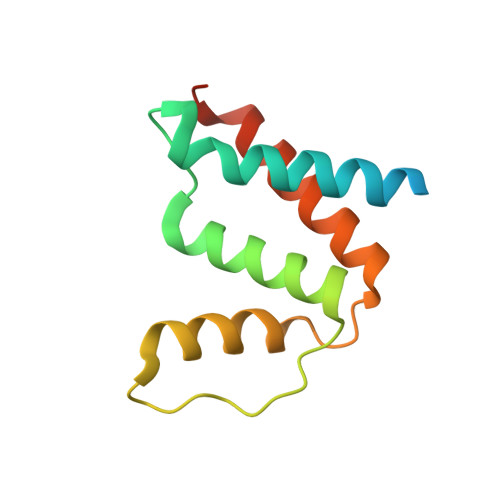 | |
UniProt & NIH Common Fund Data Resources | |||||
PHAROS: Q5T8D3 GTEx: ENSG00000107897 | |||||
Entity Groups | |||||
| Sequence Clusters | 30% Identity50% Identity70% Identity90% Identity95% Identity100% Identity | ||||
| UniProt Group | Q5T8D3 | ||||
Sequence AnnotationsExpand | |||||
Reference Sequence | |||||
| Ligands 3 Unique | |||||
|---|---|---|---|---|---|
| ID | Chains | Name / Formula / InChI Key | 2D Diagram | 3D Interactions | |
| COA Download:Ideal Coordinates CCD File | D [auth A], E [auth B] | COENZYME A C21 H36 N7 O16 P3 S RGJOEKWQDUBAIZ-IBOSZNHHSA-N |  | ||
| STE Download:Ideal Coordinates CCD File | C [auth A] | STEARIC ACID C18 H36 O2 QIQXTHQIDYTFRH-UHFFFAOYSA-N |  | ||
| UNX Download:Ideal Coordinates CCD File | F [auth B], G [auth B], H [auth B], I [auth B] | UNKNOWN ATOM OR ION X |  | ||
| Length ( Å ) | Angle ( ˚ ) |
|---|---|
| a = 136.751 | α = 90 |
| b = 34.064 | β = 102.16 |
| c = 54.877 | γ = 90 |
| Software Name | Purpose |
|---|---|
| MAR345 | data collection |
| PHASER | phasing |
| REFMAC | refinement |
| MOSFLM | data reduction |
| SCALA | data scaling |


