


Structural and Site-directed Mutagenesis Study of Versatile Peroxidase Oxidizing both Mn(II) and Aromatic Substrates
Piontek, K., Choinowski, T., Perez-Boada, M., Ruiz-Duenas, F.J., Martinez, M.J., Plattner, D.A., Martinez, A.T.To be published.
Experimental Data Snapshot
Starting Model: experimental
View more details
Entity ID: 1 | |||||
|---|---|---|---|---|---|
| Molecule | Chains | Sequence Length | Organism | Details | Image |
| Versatile peroxidase VPL2 | 331 | Pleurotus eryngii | Mutation(s): 0 EC: 1.11.1.16 | 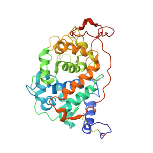 | |
UniProt | |||||
Entity Groups | |||||
| Sequence Clusters | 30% Identity50% Identity70% Identity90% Identity95% Identity100% Identity | ||||
| UniProt Group | O94753 | ||||
Glycosylation | |||||
| Glycosylation Sites: 5 | |||||
Sequence AnnotationsExpand | |||||
Reference Sequence | |||||
| Ligands 5 Unique | |||||
|---|---|---|---|---|---|
| ID | Chains | Name / Formula / InChI Key | 2D Diagram | 3D Interactions | |
| HEM Download:Ideal Coordinates CCD File | E [auth A], M [auth B] | PROTOPORPHYRIN IX CONTAINING FE C34 H32 Fe N4 O4 KABFMIBPWCXCRK-RGGAHWMASA-L |  | ||
| NAG Download:Ideal Coordinates CCD File | I [auth A], Q [auth B] | 2-acetamido-2-deoxy-beta-D-glucopyranose C8 H15 N O6 OVRNDRQMDRJTHS-FMDGEEDCSA-N |  | ||
| MAN Download:Ideal Coordinates CCD File | J [auth A] K [auth A] L [auth A] R [auth B] S [auth B] | alpha-D-mannopyranose C6 H12 O6 WQZGKKKJIJFFOK-PQMKYFCFSA-N |  | ||
| MN Download:Ideal Coordinates CCD File | H [auth A], P [auth B] | MANGANESE (II) ION Mn WAEMQWOKJMHJLA-UHFFFAOYSA-N |  | ||
| CA Download:Ideal Coordinates CCD File | F [auth A], G [auth A], N [auth B], O [auth B] | CALCIUM ION Ca BHPQYMZQTOCNFJ-UHFFFAOYSA-N |  | ||
| Length ( Å ) | Angle ( ˚ ) |
|---|---|
| a = 73.141 | α = 90 |
| b = 92.762 | β = 90 |
| c = 113.278 | γ = 90 |
| Software Name | Purpose |
|---|---|
| REFMAC | refinement |


