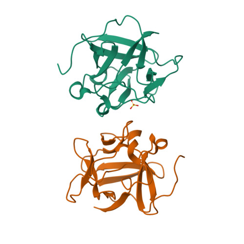
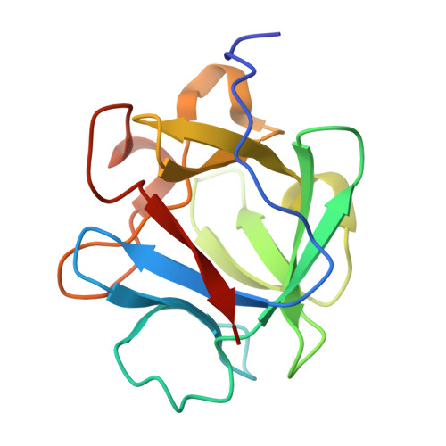
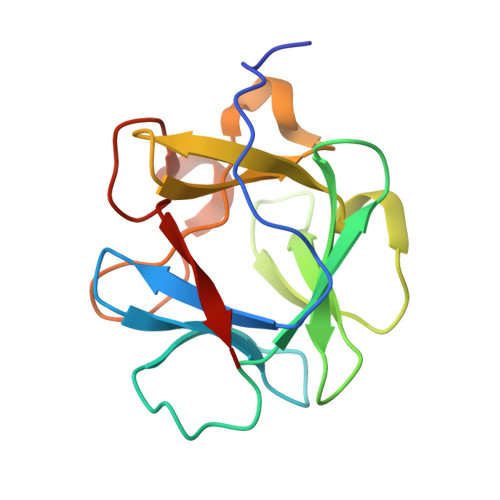
3FJD

Crystal structure of L44F/F132W mutant of Human acidic fibroblast growth factor


Experimental Data Snapshot

  • Method: X-RAY DIFFRACTION
  • Resolution: 1.90 Å
  • R-Value Free: 0.221 
  • R-Value Work: 0.190 
  • R-Value Observed: 0.190 

Starting Model: experimental
View more details

wwPDB Validation   3D Report Full Report


This is version 1.3 of the entry. See complete history


Literature

The interaction between thermodynamic stability and buried free cysteines in regulating the functional half-life of fibroblast growth factor-1.

Lee, J.Blaber, M.

(2009) J Mol Biol 393: 113-127

  • DOI: https://doi.org/10.1016/j.jmb.2009.08.026
  • Primary Citation of Related Structures:  
    3FGM, 3FJ8, 3FJ9, 3FJA, 3FJB, 3FJC, 3FJD

  • PubMed Abstract: 

    Protein biopharmaceuticals are an important and growing area of human therapeutics; however, the intrinsic property of proteins to adopt alternative conformations (such as during protein unfolding and aggregation) presents numerous challenges, limiting their effective application as biopharmaceuticals. Using fibroblast growth factor-1 as model system, we describe a cooperative interaction between the intrinsic property of thermostability and the reactivity of buried free-cysteine residues that can substantially modulate protein functional half-life. A mutational strategy that combines elimination of buried free cysteines and secondary mutations that enhance thermostability to achieve a substantial gain in functional half-life is described. Furthermore, the implementation of this design strategy utilizing stabilizing mutations within the core region resulted in a mutant protein that is essentially indistinguishable from wild type as regard protein surface and solvent structure, thus minimizing the immunogenic potential of the mutations. This design strategy should be generally applicable to soluble globular proteins containing buried free-cysteine residues.


  • Organizational Affiliation

    Department of Biomedical Sciences, College of Medicine, Florida State University, Tallahassee, FL 32306-4300, USA.


Macromolecules
Find similar proteins by:  (by identity cutoff)  |  3D Structure
Entity ID: 1
MoleculeChains Sequence LengthOrganismDetailsImage
Heparin-binding growth factor 1
A, B
146Homo sapiensMutation(s): 2 
Gene Names: FGF1FGFA
UniProt & NIH Common Fund Data Resources
Find proteins for P05230 (Homo sapiens)
Explore P05230 
Go to UniProtKB:  P05230
PHAROS:  P05230
GTEx:  ENSG00000113578 
Entity Groups  
Sequence Clusters30% Identity50% Identity70% Identity90% Identity95% Identity100% Identity
UniProt GroupP05230
Sequence Annotations
Expand
  • Reference Sequence
Experimental Data & Validation

Experimental Data

  • Method: X-RAY DIFFRACTION
  • Resolution: 1.90 Å
  • R-Value Free: 0.221 
  • R-Value Work: 0.190 
  • R-Value Observed: 0.190 
  • Space Group: C 2 2 21
Unit Cell:
Length ( Å )Angle ( ˚ )
a = 73.236α = 90
b = 97.581β = 90
c = 108.451γ = 90
Software Package:
Software NamePurpose
CrystalCleardata collection
CNSrefinement
DENZOdata reduction
SCALEPACKdata scaling
CNSphasing

Structure Validation

View Full Validation Report


Entry History 

Deposition Data

Revision History  (Full details and data files)

  • Version 1.0: 2009-10-06
    Type: Initial release
  • Version 1.1: 2011-07-13
    Changes: Version format compliance
  • Version 1.2: 2021-10-20
    Changes: Database references, Derived calculations
  • Version 1.3: 2023-09-06
    Changes: Data collection, Refinement description