3FJ5 | pdb_00003fj5

Crystal structure of the c-src-SH3 domain


Experimental Data Snapshot

  • Method: X-RAY DIFFRACTION
  • Resolution: 1.65 Å
  • R-Value Free: 
    0.222 (Depositor), 0.265 (DCC) 
  • R-Value Work: 
    0.206 (Depositor), 0.266 (DCC) 
  • R-Value Observed: 
    0.206 (Depositor) 

Starting Model: experimental
View more details

wwPDB Validation 3D Report Full Report

Validation slider image for 3FJ5

This is version 1.3 of the entry. See complete history

Literature

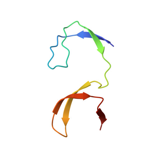
Intertwined dimeric structure for the SH3 domain of the c-Src tyrosine kinase induced by polyethylene glycol binding

Morel, B.Ruiz-Sanz, J.Luque, I.

(2009) FEBS Lett 583: 749-753

  • DOI: https://doi.org/10.1016/j.febslet.2009.01.036
  • Primary Citation Related Structures: 
    3FJ5

  • PubMed Abstract: 

    Here we report the first crystal structure of the SH3 domain of the cellular Src tyrosine kinase (c-Src-SH3) domain on its own. In the crystal two molecules of c-Src-SH3 exchange their -RT loops generating an intertwined dimer, in which the two SH3 units, preserving the binding site configuration, are oriented to allow simultaneous binding of two ligand molecules. The dimerization of c-Src-SH3 is induced, both in the crystal and in solution, by the binding of a PEG molecule at the dimer interface, indicating that this type of conformations are energetically close to the native structure. These results have important implications respect to in vivo oligomerization and amyloid aggregation.


  • Organizational Affiliation
    • Department of Physical Chemistry, Biochemistry and Inorganic Chemistry, University of Almería, Carretera Sacramento s/n, 04120 Almería, Spain.

Macromolecule Content 

  • Total Structure Weight: 13.98 kDa 
  • Atom Count: 1,036 
  • Modeled Residue Count: 114 
  • Deposited Residue Count: 114 
  • Unique protein chains: 1

Macromolecules

Find similar proteins by:|  3D Structure
Entity ID: 1
MoleculeChains  Sequence LengthOrganismDetailsImage
Proto-oncogene tyrosine-protein kinase Src
A, B
57Gallus gallusMutation(s): 1 
Gene Names: c-SRCSRC
EC: 2.7.10.2
UniProt
Find proteins for P00523 (Gallus gallus)
Explore P00523 
Go to UniProtKB:  P00523
Entity Groups
Sequence Clusters30% Identity50% Identity70% Identity90% Identity95% Identity100% Identity
UniProt GroupP00523
Sequence Annotations
Expand
Reference Sequence

Small Molecules

Ligands 5 Unique
IDChains Name / Formula / InChI Key2D Diagram3D Interactions
PG4

Query on PG4


Download:Ideal Coordinates CCD File
G [auth A],
J [auth B]
TETRAETHYLENE GLYCOL
C8 H18 O5
UWHCKJMYHZGTIT-UHFFFAOYSA-N
PGE

Query on PGE


Download:Ideal Coordinates CCD File
C [auth A]TRIETHYLENE GLYCOL
C6 H14 O4
ZIBGPFATKBEMQZ-UHFFFAOYSA-N
SO4

Query on SO4


Download:Ideal Coordinates CCD File
D [auth A]SULFATE ION
O4 S
QAOWNCQODCNURD-UHFFFAOYSA-L
GOL

Query on GOL


Download:Ideal Coordinates CCD File
F [auth A]GLYCEROL
C3 H8 O3
PEDCQBHIVMGVHV-UHFFFAOYSA-N
ACT

Query on ACT


Download:Ideal Coordinates CCD File
E [auth A],
H [auth B],
I [auth B]
ACETATE ION
C2 H3 O2
QTBSBXVTEAMEQO-UHFFFAOYSA-M

Experimental Data & Validation

Experimental Data

  • Method: X-RAY DIFFRACTION
  • Resolution: 1.65 Å
  • R-Value Free:  0.222 (Depositor), 0.265 (DCC) 
  • R-Value Work:  0.206 (Depositor), 0.266 (DCC) 
  • R-Value Observed: 0.206 (Depositor) 
Space Group: P 65
Unit Cell:
Length ( Å )Angle ( ˚ )
a = 46.596α = 90
b = 46.596β = 90
c = 128.064γ = 120
Software Package:
Software NamePurpose
MOSFLMdata reduction
SCALAdata scaling
MOLREPphasing
REFMACrefinement
PDB_EXTRACTdata extraction
HKL-2000data collection

Structure Validation

View Full Validation Report


Entry History 

Deposition Data

Revision History  (Full details and data files)

  • Version 1.0: 2009-03-03
    Type: Initial release
  • Version 1.1: 2011-07-13
    Changes: Non-polymer description, Version format compliance
  • Version 1.2: 2021-11-10
    Changes: Database references, Derived calculations
  • Version 1.3: 2023-11-01
    Changes: Data collection, Refinement description