3FIU | pdb_00003fiu

Structure of NMN synthetase from Francisella tularensis


Experimental Data Snapshot

  • Method: X-RAY DIFFRACTION
  • Resolution: 1.85 Å
  • R-Value Free: 
    0.233 (Depositor), 0.234 (DCC) 
  • R-Value Work: 
    0.184 (Depositor) 
  • R-Value Observed: 
    0.187 (Depositor) 

Starting Model: experimental
View more details

wwPDB Validation 3D Report Full Report

Validation slider image for 3FIU

Ligand Structure Quality Assessment 


This is version 1.4 of the entry. See complete history

Literature

Nicotinamide mononucleotide synthetase is the key enzyme for an alternative route of NAD biosynthesis in Francisella tularensis

Sorci, L.Martynowski, D.Rodionov, D.A.Eyobo, Y.Zogaj, X.Klose, K.E.Nikolaev, E.V.Magni, G.Zhang, H.Osterman, A.L.

(2009) Proc Natl Acad Sci U S A 106: 3083-3088

  • DOI: https://doi.org/10.1073/pnas.0811718106
  • Primary Citation Related Structures: 
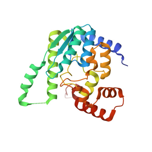
    3FIU

  • PubMed Abstract: 

    Enzymes involved in the last 2 steps of nicotinamide adenine dinucleotide (NAD) cofactor biosynthesis, which catalyze the adenylylation of the nicotinic acid mononucleotide (NaMN) precursor to nicotinic acid dinucleotide (NaAD) followed by its amidation to NAD, constitute promising drug targets for the development of new antibiotics. These enzymes, NaMN adenylyltransferase (gene nadD) and NAD synthetase (gene nadE), respectively, are indispensable and conserved in nearly all bacterial pathogens. However, a comparative genome analysis of Francisella tularensis allowed us to predict the existence of an alternative route of NAD synthesis in this category A priority pathogen, the causative agent of tularaemia. In this route, the amidation of NaMN to nicotinamide mononucleotide (NMN) occurs before the adenylylation reaction, which converts this alternative intermediate to the NAD cofactor. The first step is catalyzed by NMN synthetase, which was identified and characterized in this study. A crystal structure of this enzyme, a divergent member of the NadE family, was solved at 1.9-A resolution in complex with reaction products, providing a rationale for its unusual substrate preference for NaMN over NaAD. The second step is performed by NMN adenylyltransferase of the NadM family. Here, we report validation of the predicted route (NaMN --> NMN --> NAD) in F. tularensis including mathematical modeling, in vitro reconstitution, and in vivo metabolite analysis in comparison with a canonical route (NaMN --> NaAD --> NAD) of NAD biosynthesis as represented by another deadly bacterial pathogen, Bacillus anthracis.


  • Organizational Affiliation
    • Burnham Institute for Medical Research, 10901 North Torrey Pines Road, La Jolla, CA 92037, USA.

Macromolecule Content 

  • Total Structure Weight: 114.83 kDa 
  • Atom Count: 8,516 
  • Modeled Residue Count: 952 
  • Deposited Residue Count: 996 
  • Unique protein chains: 1

Macromolecules

Find similar proteins by:|  3D Structure
Entity ID: 1
MoleculeChains  Sequence LengthOrganismDetailsImage
NH(3)-dependent NAD(+) synthetase
A, B, C, D
249Francisella tularensis subsp. holarctica LVSMutation(s): 0 
Gene Names: FTL_0685NadE
EC: 6.3.1.5

Small Molecules

Ligands 4 Unique
IDChains Name / Formula / InChI Key2D Diagram3D Interactions
AMP

Query on AMP


Download:Ideal Coordinates CCD File
G [auth A],
L [auth B],
Q [auth C],
V [auth D]
ADENOSINE MONOPHOSPHATE
C10 H14 N5 O7 P
UDMBCSSLTHHNCD-KQYNXXCUSA-N
POP

Query on POP


Download:Ideal Coordinates CCD File
H [auth A],
M [auth B],
R [auth C],
W [auth D]
PYROPHOSPHATE 2-
H2 O7 P2
XPPKVPWEQAFLFU-UHFFFAOYSA-L
MG

Query on MG


Download:Ideal Coordinates CCD File
E [auth A]
F [auth A]
J [auth B]
K [auth B]
O [auth C]
E [auth A],
F [auth A],
J [auth B],
K [auth B],
O [auth C],
P [auth C],
T [auth D],
U [auth D]
MAGNESIUM ION
Mg
JLVVSXFLKOJNIY-UHFFFAOYSA-N
NA

Query on NA


Download:Ideal Coordinates CCD File
I [auth A],
N [auth B],
S [auth C],
X [auth D]
SODIUM ION
Na
FKNQFGJONOIPTF-UHFFFAOYSA-N

Experimental Data & Validation

Experimental Data

  • Method: X-RAY DIFFRACTION
  • Resolution: 1.85 Å
  • R-Value Free:  0.233 (Depositor), 0.234 (DCC) 
  • R-Value Work:  0.184 (Depositor) 
  • R-Value Observed: 0.187 (Depositor) 
Space Group: P 21 21 21
Unit Cell:
Length ( Å )Angle ( ˚ )
a = 50.432α = 90
b = 126.438β = 90
c = 152.531γ = 90
Software Package:
Software NamePurpose
HKL-2000data collection
MOLREPphasing
REFMACrefinement
HKL-2000data reduction
HKL-2000data scaling

Structure Validation

View Full Validation Report


Ligand Structure Quality Assessment 


Entry History 

Deposition Data

Revision History  (Full details and data files)

  • Version 1.0: 2009-03-03
    Type: Initial release
  • Version 1.1: 2011-07-13
    Changes: Version format compliance
  • Version 1.2: 2014-11-12
    Changes: Structure summary
  • Version 1.3: 2017-11-01
    Changes: Refinement description
  • Version 1.4: 2023-09-06
    Changes: Data collection, Database references, Derived calculations, Refinement description