3FHO | pdb_00003fho

Structure of S. pombe Dbp5


Experimental Data Snapshot

  • Method: X-RAY DIFFRACTION
  • Resolution: 2.80 Å
  • R-Value Free: 
    0.327 (Depositor), 0.326 (DCC) 
  • R-Value Work: 
    0.290 (Depositor), 0.291 (DCC) 
  • R-Value Observed: 
    0.292 (Depositor) 

Starting Model: experimental
View more details

wwPDB Validation 3D Report Full Report

Validation slider image for 3FHO

This is version 1.2 of the entry. See complete history

Literature

Solution and crystal structures of mRNA exporter Dbp5p and its interaction with nucleotides

Fan, J.S.Cheng, Z.Zhang, J.Noble, C.Zhou, Z.Song, H.Yang, D.

(2009) J Mol Biology 388: 1-10

  • DOI: https://doi.org/10.1016/j.jmb.2009.03.004
  • Primary Citation Related Structures: 
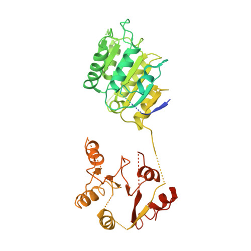
    2KBE, 2KBF, 3FHO

  • PubMed Abstract: 

    DEAD-box protein 5 (Dbp5p) plays very important roles in RNA metabolism from transcription, to translation, to RNA decay. It is an RNA helicase and functions as an essential RNA export factor from nucleus. Here, we report the solution NMR structures of the N- and C-terminal domains (NTD and CTD, respectively) of Dbp5p from Saccharomyces cerevisiae (ScDbp5p) and X-ray crystal structure of Dbp5p from Schizosaccharomyces pombe (SpDbp5p) in the absence of nucleotides and RNA. The crystal structure clearly shows that SpDbp5p comprises two RecA-like domains that do not interact with each other. NMR results show that the N-terminal flanking region of ScDpbp5 (M1-E70) is intrinsically unstructured and the region Y71-R121 including the Q motif is highly dynamic on millisecond-microsecond timescales in solution. The C-terminal flanking region of ScDbp5p forms a short beta-strand and a long helix. This helix is unique for ScDbp5p and has not been observed in other DEAD-box proteins. Compared with other DEAD-box proteins, Dbp5p has an extra insert with six residues in the CTD. NMR structure reveals that the insert is located in a solvent-exposed loop capable of interacting with other proteins. ATP and ADP titration experiments show that both ADP and ATP bind to the consensus binding site in the NTD of ScDbp5p but do not interact with the CTD at all. Binding of ATP or ADP to NTD induces significant conformational rearrangement too.


  • Organizational Affiliation
    • Department of Biological Sciences, National University of Singapore, 14 Science Drive 4, Singapore 117543, Singapore.

Macromolecule Content 

  • Total Structure Weight: 106.18 kDa 
  • Atom Count: 4,771 
  • Modeled Residue Count: 611 
  • Deposited Residue Count: 1,016 
  • Unique protein chains: 1

Macromolecules

Find similar proteins by:|  3D Structure
Entity ID: 1
MoleculeChains  Sequence LengthOrganismDetailsImage
ATP-dependent RNA helicase dbp5
A, B
508Schizosaccharomyces pombeMutation(s): 0 
Gene Names: dbp5
EC: 3.6.1 (PDB Primary Data), 3.6.4.13 (UniProt)
UniProt
Find proteins for Q09747 (Schizosaccharomyces pombe (strain 972 / ATCC 24843))
Explore Q09747 
Go to UniProtKB:  Q09747
Entity Groups
Sequence Clusters30% Identity50% Identity70% Identity90% Identity95% Identity100% Identity
UniProt GroupQ09747
Sequence Annotations
Expand
Reference Sequence

Experimental Data & Validation

Experimental Data

  • Method: X-RAY DIFFRACTION
  • Resolution: 2.80 Å
  • R-Value Free:  0.327 (Depositor), 0.326 (DCC) 
  • R-Value Work:  0.290 (Depositor), 0.291 (DCC) 
  • R-Value Observed: 0.292 (Depositor) 
Space Group: C 1 2 1
Unit Cell:
Length ( Å )Angle ( ˚ )
a = 108.736α = 90
b = 144.044β = 89.96
c = 79.137γ = 90
Software Package:
Software NamePurpose
DNAdata collection
PHASERphasing
REFMACrefinement
MOSFLMdata reduction
SCALAdata scaling

Structure Validation

View Full Validation Report


Entry History 

Deposition Data

Revision History  (Full details and data files)

  • Version 1.0: 2009-10-13
    Type: Initial release
  • Version 1.1: 2011-07-13
    Changes: Advisory, Refinement description, Version format compliance
  • Version 1.2: 2023-11-01
    Changes: Data collection, Database references, Refinement description