3FHN | pdb_00003fhn

Structure of Tip20p


Experimental Data Snapshot

  • Method: X-RAY DIFFRACTION
  • Resolution: 3.00 Å
  • R-Value Free: 
    0.265 (Depositor), 0.265 (DCC) 
  • R-Value Work: 
    0.220 (Depositor), 0.220 (DCC) 
  • R-Value Observed: 
    0.220 (Depositor) 

wwPDB Validation 3D Report Full Report

Validation slider image for 3FHN

This is version 1.2 of the entry. See complete history

Literature

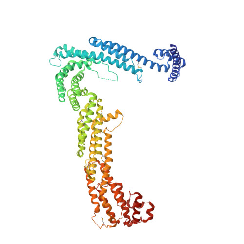
Structural characterization of Tip20p and Dsl1p, subunits of the Dsl1p vesicle tethering complex.

Tripathi, A.Ren, Y.Jeffrey, P.D.Hughson, F.M.

(2009) Nat Struct Mol Biol 16: 114-123

  • DOI: https://doi.org/10.1038/nsmb.1548
  • Primary Citation Related Structures: 
    3ETU, 3ETV, 3FHN

  • PubMed Abstract: 

    Multisubunit tethering complexes are essential for intracellular trafficking and have been proposed to mediate the initial interaction between vesicles and the membranes with which they fuse. Here we report initial structural characterization of the Dsl1p complex, whose three subunits are essential for trafficking from the Golgi apparatus to the endoplasmic reticulum (ER). Crystal structures reveal that two of the three subunits, Tip20p and Dsl1p, resemble known subunits of the exocyst complex, establishing a structural connection among several multisubunit tethering complexes and implying that many of their subunits are derived from a common progenitor. We show, moreover, that Tip20p and Dsl1p interact directly via N-terminal alpha-helices. Finally, we establish that different Dsl1p complex subunits bind independently to different ER SNARE proteins. Our results map out two alternative protein-interaction networks capable of tethering COPI-coated vesicles, via the Dsl1p complex, to ER membranes.


  • Organizational Affiliation
    • Department of Molecular Biology, Princeton University, Princeton, New Jersey 08544, USA.

Macromolecule Content 

  • Total Structure Weight: 328.88 kDa 
  • Atom Count: 22,040 
  • Modeled Residue Count: 2,692 
  • Deposited Residue Count: 2,824 
  • Unique protein chains: 1

Macromolecules

Find similar proteins by:|  3D Structure
Entity ID: 1
MoleculeChains  Sequence LengthOrganismDetailsImage
Protein transport protein TIP20
A, B, C, D
706Saccharomyces cerevisiaeMutation(s): 0 
Gene Names: TIP20TIP1YGL145W
UniProt
Find proteins for P33891 (Saccharomyces cerevisiae (strain ATCC 204508 / S288c))
Explore P33891 
Go to UniProtKB:  P33891
Entity Groups
Sequence Clusters30% Identity50% Identity70% Identity90% Identity95% Identity100% Identity
UniProt GroupP33891
Sequence Annotations
Expand
Reference Sequence

Small Molecules

Modified Residues  1 Unique
IDChains TypeFormula2D DiagramParent
MSE
Query on MSE
A, B, C, D
L-PEPTIDE LINKINGC5 H11 N O2 SeMET

Experimental Data & Validation

Experimental Data

  • Method: X-RAY DIFFRACTION
  • Resolution: 3.00 Å
  • R-Value Free:  0.265 (Depositor), 0.265 (DCC) 
  • R-Value Work:  0.220 (Depositor), 0.220 (DCC) 
  • R-Value Observed: 0.220 (Depositor) 
Space Group: P 1
Unit Cell:
Length ( Å )Angle ( ˚ )
a = 85.48α = 77.09
b = 111.62β = 88.12
c = 149.84γ = 70.35
Software Package:
Software NamePurpose
CBASSdata collection
SHELXSphasing
CNSrefinement
DENZOdata reduction
SCALEPACKdata scaling

Structure Validation

View Full Validation Report


Entry History 

Deposition Data

Revision History  (Full details and data files)

  • Version 1.0: 2009-01-20
    Type: Initial release
  • Version 1.1: 2011-07-13
    Changes: Version format compliance
  • Version 1.2: 2024-11-20
    Changes: Data collection, Database references, Derived calculations, Structure summary