3FCA | pdb_00003fca

Genetic Incorporation of a Metal-ion Chelating Amino Acid into proteins as biophysical probe


Experimental Data Snapshot

  • Method: X-RAY DIFFRACTION
  • Resolution: 2.15 Å
  • R-Value Free: 
    0.244 (Depositor), 0.241 (DCC) 
  • R-Value Work: 
    0.207 (Depositor), 0.206 (DCC) 
  • R-Value Observed: 
    0.209 (Depositor) 

wwPDB Validation 3D Report Full Report

Validation slider image for 3FCA

This is version 1.4 of the entry. See complete history

Literature

Genetic incorporation of a metal-ion chelating amino acid into proteins as a biophysical probe.

Lee, H.S.Spraggon, G.Schultz, P.G.Wang, F.

(2009) J Am Chem Soc 131: 2481-2483

  • DOI: https://doi.org/10.1021/ja808340b
  • Primary Citation Related Structures: 
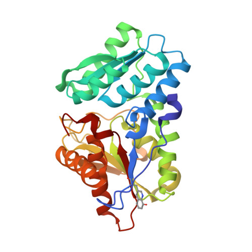
    3FCA

  • PubMed Abstract: 

    A metal-ion chelating amino acid, (8-hydroxyquinolin-3-yl)alanine, was genetically encoded in E. coli by an amber nonsense codon and corresponding orthogonal tRNA/aminoacyl-tRNA synthetase pair. The amino acid was incorporated into TM0665 protein, and the mutant protein was cocrystallized with Zn(2+) to determine the structure by SAD phasing. The structure showed a high occupancy of the heavy metal bound to the HQ-Ala residue, and the heavy metal provided excellent phasing power to determine the structure. This method also facilitates the de novo design of metalloproteins with novel structures and functions, including fluorescent sensors.


  • Organizational Affiliation
    • Department of Chemistry, The Scripps Research Institute, 10550 North Torrey Pines Road, La Jolla, California 92037, USA.

Macromolecule Content 

  • Total Structure Weight: 62.61 kDa 
  • Atom Count: 4,542 
  • Modeled Residue Count: 582 
  • Deposited Residue Count: 582 
  • Unique protein chains: 1

Macromolecules

Find similar proteins by:|  3D Structure
Entity ID: 1
MoleculeChains  Sequence LengthOrganismDetailsImage
Cysteine synthase
A, B
291Thermus thermophilusMutation(s): 0 
EC: 2.5.1.47

Small Molecules

Modified Residues  1 Unique
IDChains TypeFormula2D DiagramParent
HQA
Query on HQA
A, B
L-PEPTIDE LINKINGC12 H12 N2 O3ALA

Experimental Data & Validation

Experimental Data

  • Method: X-RAY DIFFRACTION
  • Resolution: 2.15 Å
  • R-Value Free:  0.244 (Depositor), 0.241 (DCC) 
  • R-Value Work:  0.207 (Depositor), 0.206 (DCC) 
  • R-Value Observed: 0.209 (Depositor) 
Space Group: P 42 21 2
Unit Cell:
Length ( Å )Angle ( ˚ )
a = 135.858α = 90
b = 135.858β = 90
c = 74.939γ = 90
Software Package:
Software NamePurpose
DENZOdata reduction
SCALEPACKdata scaling
PHASESphasing
RESOLVEphasing
REFMACrefinement
PDB_EXTRACTdata extraction
HKL-2000data collection

Structure Validation

View Full Validation Report


Entry History 

Deposition Data

Revision History  (Full details and data files)

  • Version 1.0: 2009-02-17
    Type: Initial release
  • Version 1.1: 2011-07-13
    Changes: Version format compliance
  • Version 1.2: 2017-11-01
    Changes: Refinement description
  • Version 1.3: 2023-12-27
    Changes: Data collection, Database references, Derived calculations
  • Version 1.4: 2024-03-27
    Changes: Derived calculations