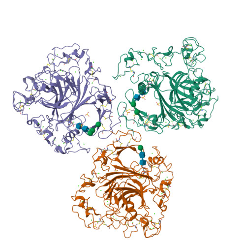
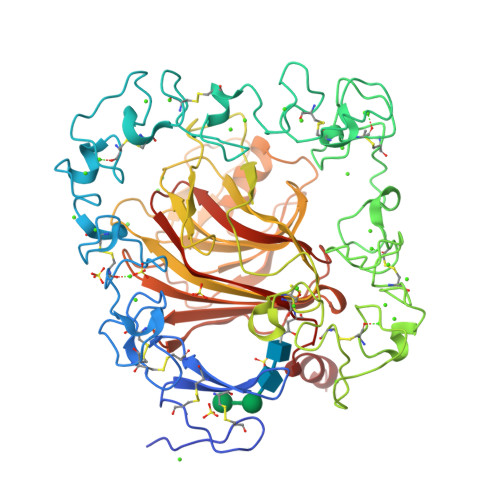
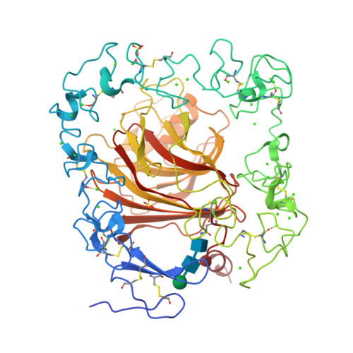
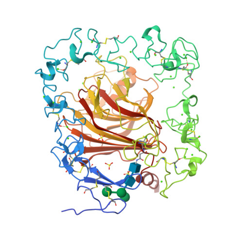
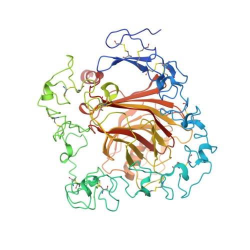
3FBY

The crystal structure of the signature domain of cartilage oligomeric matrix protein.


Experimental Data Snapshot

  • Method: X-RAY DIFFRACTION
  • Resolution: 3.15 Å
  • R-Value Free: 0.266 
  • R-Value Work: 0.202 
  • R-Value Observed: 0.205 

Starting Models: experimental
View more details

wwPDB Validation   3D Report Full Report


This is version 2.2 of the entry. See complete history


Literature

The crystal structure of the signature domain of cartilage oligomeric matrix protein: implications for collagen, glycosaminoglycan and integrin binding.

Tan, K.Duquette, M.Joachimiak, A.Lawler, J.

(2009) FASEB J 23: 2490-2501

  • DOI: https://doi.org/10.1096/fj.08-128090
  • Primary Citation of Related Structures:  
    3FBY

  • PubMed Abstract: 

    Cartilage oligomeric matrix protein (COMP), or thrombospondin-5 (TSP-5), is a secreted glycoprotein that is important for growth plate organization and function. Mutations in COMP cause two skeletal dysplasias, pseudoachondroplasia (PSACH) and multiple epiphyseal dysplasia (EDM1). In this study, we determined the structure of a recombinant protein that contains the last epidermal growth factor repeat, the type 3 repeats and the C-terminal domain (CTD) of COMP to 3.15-A resolution limit by X-ray crystallography. The CTD is a beta-sandwich that is composed of 15 antiparallel beta-strands, and the type 3 repeats are a contiguous series of calcium binding sites that associate with the CTD at multiple points. The crystal packing reveals an exposed potential metal-ion-dependent adhesion site (MIDAS) on one edge of the beta-sandwich that is common to all TSPs and may serve as a binding site for collagens and other ligands. Disease-causing mutations in COMP disrupt calcium binding, disulfide bond formation, intramolecular interactions, or sites for potential ligand binding. The structure presented here and its unique molecular packing in the crystal identify potential interactive sites for glycosaminoglycans, integrins, and collagens, which are key to cartilage structure and function.


  • Organizational Affiliation

    Midwest Center for Structural Genomics, Biosciences Division, Argonne National Laboratory, Argonne, Illinois, USA.


Macromolecules
Find similar proteins by:  (by identity cutoff)  |  3D Structure
Entity ID: 1
MoleculeChains Sequence LengthOrganismDetailsImage
Cartilage oligomeric matrix protein
A, B, C
551Homo sapiensMutation(s): 0 
Gene Names: COMPCOMP(cartilage oligomeric matrix protein)
UniProt & NIH Common Fund Data Resources
Find proteins for P49747 (Homo sapiens)
Explore P49747 
Go to UniProtKB:  P49747
PHAROS:  P49747
GTEx:  ENSG00000105664 
Entity Groups  
Sequence Clusters30% Identity50% Identity70% Identity90% Identity95% Identity100% Identity
UniProt GroupP49747
Glycosylation
Glycosylation Sites: 1Go to GlyGen: P49747-1
Sequence Annotations
Expand
  • Reference Sequence
Oligosaccharides

Help

Entity ID: 2
MoleculeChains Length2D Diagram Glycosylation3D Interactions
alpha-D-mannopyranose-(1-6)-alpha-D-mannopyranose-(1-4)-2-acetamido-2-deoxy-beta-D-glucopyranose-(1-4)-2-acetamido-2-deoxy-beta-D-glucopyranose
D
4N-Glycosylation
Glycosylation Resources
GlyTouCan:  G04854NQ
GlyCosmos:  G04854NQ
GlyGen:  G04854NQ
Entity ID: 3
MoleculeChains Length2D Diagram Glycosylation3D Interactions
alpha-D-mannopyranose-(1-4)-2-acetamido-2-deoxy-beta-D-glucopyranose-(1-4)-2-acetamido-2-deoxy-beta-D-glucopyranose
E
3N-Glycosylation
Glycosylation Resources
GlyTouCan:  G62182OO
GlyCosmos:  G62182OO
GlyGen:  G62182OO
Entity ID: 4
MoleculeChains Length2D Diagram Glycosylation3D Interactions
alpha-D-mannopyranose-(1-3)-[alpha-D-mannopyranose-(1-6)]alpha-D-mannopyranose-(1-4)-2-acetamido-2-deoxy-beta-D-glucopyranose-(1-4)-2-acetamido-2-deoxy-beta-D-glucopyranose
F
5N-Glycosylation
Glycosylation Resources
GlyTouCan:  G21381MC
GlyCosmos:  G21381MC
GlyGen:  G21381MC
Small Molecules
Ligands 2 Unique
IDChains Name / Formula / InChI Key2D Diagram3D Interactions
SO4
Query on SO4

Download Ideal Coordinates CCD File 
KA [auth A]
LA [auth A]
MA [auth A]
NA [auth A]
OA [auth A]
KA [auth A],
LA [auth A],
MA [auth A],
NA [auth A],
OA [auth A],
SB [auth B],
TB [auth B],
XC [auth C],
YC [auth C],
ZC [auth C]
SULFATE ION
O4 S
QAOWNCQODCNURD-UHFFFAOYSA-L
CA
Query on CA

Download Ideal Coordinates CCD File 
AA [auth A]
AB [auth B]
AC [auth C]
BA [auth A]
BB [auth B]
AA [auth A],
AB [auth B],
AC [auth C],
BA [auth A],
BB [auth B],
BC [auth C],
CA [auth A],
CB [auth B],
CC [auth C],
DA [auth A],
DB [auth B],
DC [auth C],
EA [auth A],
EB [auth B],
EC [auth C],
FA [auth A],
FB [auth B],
FC [auth C],
G [auth A],
GA [auth A],
GB [auth B],
GC [auth C],
H [auth A],
HA [auth A],
HB [auth B],
HC [auth C],
I [auth A],
IA [auth A],
IB [auth B],
IC [auth C],
J [auth A],
JA [auth A],
JB [auth B],
JC [auth C],
K [auth A],
KB [auth B],
KC [auth C],
L [auth A],
LB [auth B],
LC [auth C],
M [auth A],
MB [auth B],
MC [auth C],
N [auth A],
NB [auth B],
NC [auth C],
O [auth A],
OB [auth B],
OC [auth C],
P [auth A],
PA [auth B],
PB [auth B],
PC [auth C],
Q [auth A],
QA [auth B],
QB [auth B],
QC [auth C],
R [auth A],
RA [auth B],
RB [auth B],
RC [auth C],
S [auth A],
SA [auth B],
SC [auth C],
T [auth A],
TA [auth B],
TC [auth C],
U [auth A],
UA [auth B],
UB [auth C],
UC [auth C],
V [auth A],
VA [auth B],
VB [auth C],
VC [auth C],
W [auth A],
WA [auth B],
WB [auth C],
WC [auth C],
X [auth A],
XA [auth B],
XB [auth C],
Y [auth A],
YA [auth B],
YB [auth C],
Z [auth A],
ZA [auth B],
ZB [auth C]
CALCIUM ION
Ca
BHPQYMZQTOCNFJ-UHFFFAOYSA-N
Experimental Data & Validation

Experimental Data

  • Method: X-RAY DIFFRACTION
  • Resolution: 3.15 Å
  • R-Value Free: 0.266 
  • R-Value Work: 0.202 
  • R-Value Observed: 0.205 
  • Space Group: P 32 2 1
Unit Cell:
Length ( Å )Angle ( ˚ )
a = 192.242α = 90
b = 192.242β = 90
c = 145.681γ = 120
Software Package:
Software NamePurpose
SBC-Collectdata collection
MOLREPphasing
REFMACrefinement
HKL-3000data reduction
HKL-3000data scaling

Structure Validation

View Full Validation Report


Entry History 

Deposition Data

Revision History  (Full details and data files)

  • Version 1.0: 2009-06-23
    Type: Initial release
  • Version 1.1: 2011-07-13
    Changes: Advisory, Version format compliance
  • Version 2.0: 2020-07-29
    Type: Remediation
    Reason: Carbohydrate remediation
    Changes: Advisory, Atomic model, Data collection, Database references, Derived calculations, Structure summary
  • Version 2.1: 2023-09-06
    Changes: Data collection, Database references, Refinement description, Structure summary
  • Version 2.2: 2024-11-20
    Changes: Structure summary