3FAW | pdb_00003faw

Crystal Structure of the Group B Streptococcus Pullulanase SAP


Experimental Data Snapshot

  • Method: X-RAY DIFFRACTION
  • Resolution: 2.10 Å
  • R-Value Free: 
    0.235 (Depositor), 0.243 (DCC) 
  • R-Value Work: 
    0.194 (Depositor), 0.200 (DCC) 
  • R-Value Observed: 
    0.196 (Depositor) 

Starting Model: experimental
View more details

wwPDB Validation 3D Report Full Report

Validation slider image for 3FAW

This is version 1.2 of the entry. See complete history

Literature

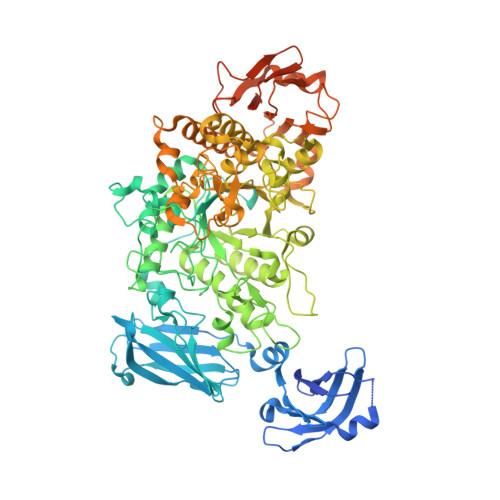
Group B Streptococcus pullulanase crystal structures in the context of a novel strategy for vaccine development

Gourlay, L.J.Santi, I.Pezzicoli, A.Grandi, G.Soriani, M.Bolognesi, M.

(2009) J Bacteriol 191: 3544-3552

  • DOI: https://doi.org/10.1128/JB.01755-08
  • Primary Citation Related Structures: 
    3FAW, 3FAX

  • PubMed Abstract: 

    The group B streptococcus type I pullulanase (SAP) is a class 13 glycoside hydrolase that is anchored to the bacterial cell surface via a conserved C-terminal anchoring motif and involved in alpha-glucan degradation. Recent in vitro functional studies have shown that SAP is immunogenic in humans and that anti-SAP sera derived from immunized animals impair both group A and group B streptococcus pullulanase activities, suggesting that in vivo immunization with this antigen could prevent streptococcal colonization. To further investigate the putative role of SAP in bacterial pathogenesis, we carried out functional studies and found that recombinant SAP binds to human cervical epithelial cells. Furthermore, with a view of using SAP as a vaccine candidate, we present high-resolution crystal structure analyses of an N-terminally truncated form of SAP lacking the carbohydrate binding module but containing the catalytic domain and displaying glycosidase hydrolase activity, both in its apo form and in complex with maltotetraose, at resolutions of 2.1 and 2.4 A, respectively.


  • Organizational Affiliation
    • Department of Biomolecular Sciences and Biotechnology, University of Milan, Via Celoria 26, I-20133 Milan, Italy.

Macromolecule Content 

  • Total Structure Weight: 98.9 kDa 
  • Atom Count: 6,593 
  • Modeled Residue Count: 775 
  • Deposited Residue Count: 877 
  • Unique protein chains: 1

Macromolecules

Find similar proteins by:|  3D Structure
Entity ID: 1
MoleculeChains  Sequence LengthOrganismDetailsImage
Reticulocyte binding protein877Streptococcus agalactiae COH1Mutation(s): 0 
Gene Names: SAN_1346

Small Molecules

Ligands 2 Unique
IDChains Name / Formula / InChI Key2D Diagram3D Interactions
CA

Query on CA


Download:Ideal Coordinates CCD File
B [auth A],
C [auth A],
D [auth A],
E [auth A]
CALCIUM ION
Ca
BHPQYMZQTOCNFJ-UHFFFAOYSA-N
CL

Query on CL


Download:Ideal Coordinates CCD File
F [auth A],
G [auth A],
H [auth A],
I [auth A],
J [auth A]
CHLORIDE ION
Cl
VEXZGXHMUGYJMC-UHFFFAOYSA-M

Experimental Data & Validation

Experimental Data

  • Method: X-RAY DIFFRACTION
  • Resolution: 2.10 Å
  • R-Value Free:  0.235 (Depositor), 0.243 (DCC) 
  • R-Value Work:  0.194 (Depositor), 0.200 (DCC) 
  • R-Value Observed: 0.196 (Depositor) 
Space Group: P 21 21 21
Unit Cell:
Length ( Å )Angle ( ˚ )
a = 48.949α = 90
b = 102.834β = 90
c = 172.438γ = 90
Software Package:
Software NamePurpose
ADSCdata collection
MOLREPphasing
REFMACrefinement
MOSFLMdata reduction
SCALAdata scaling

Structure Validation

View Full Validation Report


Entry History 

Deposition Data

Revision History  (Full details and data files)

  • Version 1.0: 2009-04-14
    Type: Initial release
  • Version 1.1: 2011-07-13
    Changes: Version format compliance
  • Version 1.2: 2023-11-01
    Changes: Data collection, Database references, Derived calculations, Refinement description