3F72 | pdb_00003f72

Crystal Structure of the Staphylococcus aureus pI258 CadC Metal Binding Site 2 Mutant


Experimental Data Snapshot

  • Method: X-RAY DIFFRACTION
  • Resolution: 2.31 Å
  • R-Value Free: 
    0.288 (Depositor), 0.231 (DCC) 
  • R-Value Work: 
    0.237 (Depositor) 
  • R-Value Observed: 
    0.240 (Depositor) 

wwPDB Validation 3D Report Full Report

Validation slider image for 3F72

This is version 1.4 of the entry. See complete history

Literature

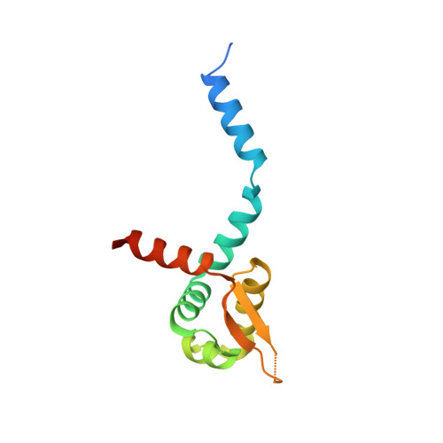
Role of bound Zn(II) in the CadC Cd(II)/Pb(II)/Zn(II)-responsive repressor.

Kandegedara, A.Thiyagarajan, S.Kondapalli, K.C.Stemmler, T.L.Rosen, B.P.

(2009) J Biological Chem 284: 14958-14965

  • DOI: https://doi.org/10.1074/jbc.M809179200
  • Primary Citation Related Structures: 
    3F72

  • PubMed Abstract: 

    The Staphylococcus aureus plasmid pI258 cadCA operon encodes a P-type ATPase, CadA, that confers resistance to Cd(II)/Pb(II)/Zn(II). Expression is regulated by CadC, a homodimeric repressor that dissociates from the cad operator/promoter upon binding of Cd(II), Pb(II), or Zn(II). CadC is a member of the ArsR/SmtB family of metalloregulatory proteins. The crystal structure of CadC shows two types of metal binding sites, termed Site 1 and Site 2, and the homodimer has two of each. Site 1 is the physiological inducer binding site. The two Site 2 metal binding sites are formed at the dimerization interface. Site 2 is not regulatory in CadC but is regulatory in the homologue SmtB. Here the role of each site was investigated by mutagenesis. Both sites bind either Cd(II) or Zn(II). However, Site 1 has higher affinity for Cd(II) over Zn(II), and Site 2 prefers Zn(II) over Cd(II). Site 2 is not required for either derepression or dimerization. The crystal structure of the wild type with bound Zn(II) and of a mutant lacking Site 2 was compared with the SmtB structure with and without bound Zn(II). We propose that an arginine residue allows for Zn(II) regulation in SmtB and, conversely, a glycine results in a lack of regulation by Zn(II) in CadC. We propose that a glycine residue was ancestral whether the repressor binds Zn(II) at a Site 2 like CadC or has no Site 2 like the paralogous ArsR and implies that acquisition of regulatory ability in SmtB was a more recent evolutionary event.


  • Organizational Affiliation
    • Department of Biochemistry and Molecular Biology, Wayne State University, School of Medicine, Detroit, Michigan 48201, USA.

Macromolecule Content 

  • Total Structure Weight: 81.89 kDa 
  • Atom Count: 4,843 
  • Modeled Residue Count: 621 
  • Deposited Residue Count: 732 
  • Unique protein chains: 1

Macromolecules

Find similar proteins by:|  3D Structure
Entity ID: 1
MoleculeChains  Sequence LengthOrganismDetailsImage
Cadmium efflux system accessory protein
A, B, C, D, E
A, B, C, D, E, F
122Staphylococcus aureusMutation(s): 3 
UniProt
Find proteins for P20047 (Staphylococcus aureus)
Explore P20047 
Go to UniProtKB:  P20047
Entity Groups
Sequence Clusters30% Identity50% Identity70% Identity90% Identity95% Identity100% Identity
UniProt GroupP20047
Sequence Annotations
Expand
Reference Sequence

Experimental Data & Validation

Experimental Data

  • Method: X-RAY DIFFRACTION
  • Resolution: 2.31 Å
  • R-Value Free:  0.288 (Depositor), 0.231 (DCC) 
  • R-Value Work:  0.237 (Depositor) 
  • R-Value Observed: 0.240 (Depositor) 
Space Group: P 43
Unit Cell:
Length ( Å )Angle ( ˚ )
a = 89.471α = 90
b = 89.471β = 90
c = 148.536γ = 90
Software Package:
Software NamePurpose
DENZOdata reduction
SCALEPACKdata scaling
PHASERphasing
REFMACrefinement
PDB_EXTRACTdata extraction
HKL-2000data collection
HKL-2000data reduction
HKL-2000data scaling

Structure Validation

View Full Validation Report


Entry History 

Deposition Data

Revision History  (Full details and data files)

  • Version 1.0: 2009-04-07
    Type: Initial release
  • Version 1.1: 2011-07-13
    Changes: Version format compliance
  • Version 1.2: 2017-10-25
    Changes: Refinement description
  • Version 1.3: 2021-10-20
    Changes: Database references, Derived calculations
  • Version 1.4: 2023-12-27
    Changes: Data collection