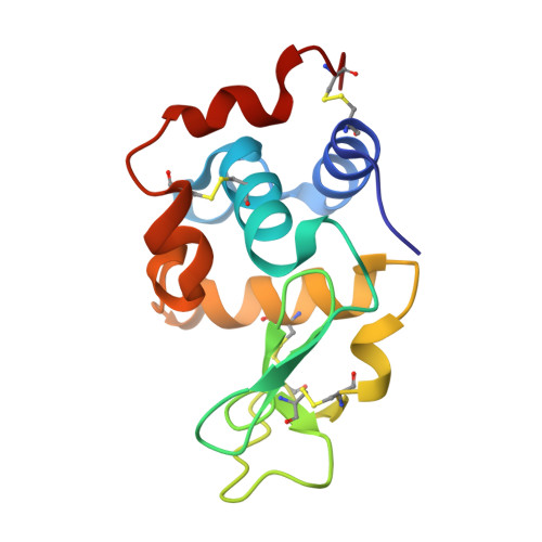
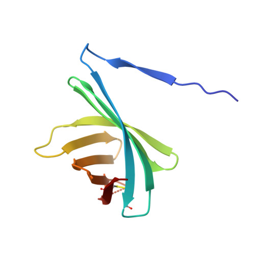
3F6Z

Crystal structure of Pseudomonas aeruginosa MliC in complex with hen egg white lysozyme


Experimental Data Snapshot

  • Method: X-RAY DIFFRACTION
  • Resolution: 2.50 Å
  • R-Value Free: 0.278 
  • R-Value Work: 0.238 
  • R-Value Observed: 0.250 

Starting Model: experimental
View more details

wwPDB Validation   3D Report Full Report


This is version 1.3 of the entry. See complete history


Literature

Structural basis for the recognition of lysozyme by MliC, a periplasmic lysozyme inhibitor in Gram-negative bacteria.

Yum, S.Kim, M.J.Xu, Y.Jin, X.L.Yoo, H.Y.Park, J.W.Gong, J.H.Choe, K.M.Lee, B.L.Ha, N.C.

(2009) Biochem Biophys Res Commun 378: 244-248

  • DOI: https://doi.org/10.1016/j.bbrc.2008.11.039
  • Primary Citation of Related Structures:  
    3F6Z

  • PubMed Abstract: 

    Lysozymes are an important component of the innate immune system of animals that hydrolyze peptidoglycan, the major bacterial cell wall constituent. Many bacteria have contrived various means of dealing with this bactericidal enzyme, one of which is to produce lysozyme inhibitors. Recently, a novel family of bacterial lysozyme inhibitors was identified in various Gram-negative bacteria, named MliC (membrane bound lysozyme inhibitor of C-type lysozyme). Here, we report the crystal structure of Pseudomonas aeruginosa MliC in complex with chicken egg white lysozyme. Combined with mutational study, the complex structure demonstrates that the invariant loop of MliC plays a crucial role in the inhibition of the lysozyme by its insertion to the active site cleft of the lysozyme, where the loop forms hydrogen and ionic bonds with the catalytic residues. Since MliC family members have been implicated as putative colonization or virulence factors, the structures and mechanism of action of MliC will be of relevance to the control of bacterial growth in animal hosts.


  • Organizational Affiliation

    College of Pharmacy and Research Institute for Drug Development, Pusan National University, Room #311, Pharmacy Building, Jangjeon-dong Geumjeong-gu, Busan 609-735, Republic of Korea.


Macromolecules
Find similar proteins by:  (by identity cutoff)  |  3D Structure
Entity ID: 1
MoleculeChains Sequence LengthOrganismDetailsImage
Lysozyme C
A, C
129Gallus gallusMutation(s): 0 
EC: 3.2.1.17
UniProt
Find proteins for P00698 (Gallus gallus)
Explore P00698 
Go to UniProtKB:  P00698
Entity Groups  
Sequence Clusters30% Identity50% Identity70% Identity90% Identity95% Identity100% Identity
UniProt GroupP00698
Sequence Annotations
Expand
  • Reference Sequence
Find similar proteins by:  (by identity cutoff)  |  3D Structure
Entity ID: 2
MoleculeChains Sequence LengthOrganismDetailsImage
Putative uncharacterized protein
B, D
101Pseudomonas aeruginosaMutation(s): 0 
Gene Names: MliCPA0867
UniProt
Find proteins for Q9I574 (Pseudomonas aeruginosa (strain ATCC 15692 / DSM 22644 / CIP 104116 / JCM 14847 / LMG 12228 / 1C / PRS 101 / PAO1))
Explore Q9I574 
Go to UniProtKB:  Q9I574
Entity Groups  
Sequence Clusters30% Identity50% Identity70% Identity90% Identity95% Identity100% Identity
UniProt GroupQ9I574
Sequence Annotations
Expand
  • Reference Sequence
Experimental Data & Validation

Experimental Data

  • Method: X-RAY DIFFRACTION
  • Resolution: 2.50 Å
  • R-Value Free: 0.278 
  • R-Value Work: 0.238 
  • R-Value Observed: 0.250 
  • Space Group: C 1 2 1
Unit Cell:
Length ( Å )Angle ( ˚ )
a = 175.97α = 90
b = 90.96β = 98.17
c = 49.73γ = 90
Software Package:
Software NamePurpose
MOSFLMdata reduction
SCALAdata scaling
MOLREPphasing
CNSrefinement
PDB_EXTRACTdata extraction
ADSCdata collection

Structure Validation

View Full Validation Report


Entry History 

Deposition Data

Revision History  (Full details and data files)

  • Version 1.0: 2008-12-23
    Type: Initial release
  • Version 1.1: 2011-07-13
    Changes: Version format compliance
  • Version 1.2: 2023-11-01
    Changes: Data collection, Database references, Refinement description
  • Version 1.3: 2024-10-30
    Changes: Structure summary