3F50 | pdb_00003f50

HIV gp41 six-helix bundle composed of an alpha/beta-peptide analogue of the CHR domain in complex with an NHR domain alpha-peptide


Experimental Data Snapshot

  • Method: X-RAY DIFFRACTION
  • Resolution: 2.80 Å
  • R-Value Free: 
    0.307 (Depositor), 0.287 (DCC) 
  • R-Value Work: 
    0.266 (Depositor) 
  • R-Value Observed: 
    0.268 (Depositor) 

Starting Models: experimental
View more details

wwPDB Validation 3D Report Full Report

Validation slider image for 3F50

This is version 2.0 of the entry. See complete history

Literature

Structural and biological mimicry of protein surface recognition by alpha/beta-peptide foldamers

Horne, W.S.Johnson, L.M.Ketas, T.J.Klasse, P.J.Lu, M.Moore, J.P.Gellman, S.H.

(2009) Proc Natl Acad Sci U S A 106: 14751-14756

  • DOI: https://doi.org/10.1073/pnas.0902663106
  • Primary Citation Related Structures: 
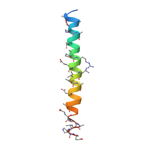
    3F4Y, 3F4Z, 3F50, 3G7A

  • PubMed Abstract: 

    Unnatural oligomers that can mimic protein surfaces offer a potentially useful strategy for blocking biomedically important protein-protein interactions. Here we evaluate an approach based on combining alpha- and beta-amino acid residues in the context of a polypeptide sequence from the HIV protein gp41, which represents an excellent testbed because of the wealth of available structural and biological information. We show that alpha/beta-peptides can mimic structural and functional properties of a critical gp41 subunit. Physical studies in solution, crystallographic data, and results from cell-fusion and virus-infectivity assays collectively indicate that the gp41-mimetic alpha/beta-peptides effectively block HIV-cell fusion via a mechanism comparable to that of gp41-derived alpha-peptides. An optimized alpha/beta-peptide is far less susceptible to proteolytic degradation than is an analogous alpha-peptide. Our findings show how a two-stage design approach, in which sequence-based alpha-->beta replacements are followed by site-specific backbone rigidification, can lead to physical and biological mimicry of a natural biorecognition process.


  • Organizational Affiliation
    • Department of Chemistry, University of Wisconsin, 1101 University Avenue, Madison, WI 53706, USA.

Macromolecule Content 

  • Total Structure Weight: 8.77 kDa 
  • Atom Count: 531 
  • Modeled Residue Count: 70 
  • Deposited Residue Count: 78 
  • Unique protein chains: 2

Macromolecules

Find similar proteins by:|  3D Structure
Entity ID: 1
MoleculeChains  Sequence LengthOrganismDetailsImage
Envelope glycoprotein gp16038N/AMutation(s): 0 
UniProt
Find proteins for P04580 (Human immunodeficiency virus type 1 group M subtype D (isolate Z6))
Explore P04580 
Go to UniProtKB:  P04580
Entity Groups
Sequence Clusters30% Identity50% Identity70% Identity90% Identity95% Identity100% Identity
UniProt GroupP04580
Sequence Annotations
Expand
Reference Sequence
Find similar proteins by:|  3D Structure
Entity ID: 2
MoleculeChains  Sequence LengthOrganismDetailsImage
alpha/beta-peptide analogue of the HIV gp41 CHR domain40N/AMutation(s): 0 

Small Molecules

Ligands 1 Unique
IDChains Name / Formula / InChI Key2D Diagram3D Interactions
GOL

Query on GOL


Download:Ideal Coordinates CCD File
C [auth A]GLYCEROL
C3 H8 O3
PEDCQBHIVMGVHV-UHFFFAOYSA-N
Modified Residues  1 Unique
IDChains TypeFormula2D DiagramParent
B3E
Query on B3E
B
L-PEPTIDE LINKINGC6 H11 N O4GLU
B3T
Query on B3T
B
L-PEPTIDE LINKINGC5 H11 N O3

--

XCP
Query on XCP
B
PEPTIDE-LIKEC6 H11 N O2

--

XPC
Query on XPC
B
PEPTIDE-LIKEC5 H10 N2 O2

--

Experimental Data & Validation

Experimental Data

  • Method: X-RAY DIFFRACTION
  • Resolution: 2.80 Å
  • R-Value Free:  0.307 (Depositor), 0.287 (DCC) 
  • R-Value Work:  0.266 (Depositor) 
  • R-Value Observed: 0.268 (Depositor) 
Space Group: P 41 3 2
Unit Cell:
Length ( Å )Angle ( ˚ )
a = 84.945α = 90
b = 84.945β = 90
c = 84.945γ = 90
Software Package:
Software NamePurpose
MD2data collection
PHASERphasing
REFMACrefinement
HKL-2000data reduction
HKL-2000data scaling

Structure Validation

View Full Validation Report


Entry History 

Deposition Data

Revision History  (Full details and data files)

  • Version 1.0: 2009-09-15
    Type: Initial release
  • Version 1.1: 2011-07-13
    Changes: Non-polymer description, Version format compliance
  • Version 1.2: 2023-09-06
    Changes: Data collection, Database references, Derived calculations, Refinement description
  • Version 2.0: 2023-11-15
    Changes: Atomic model, Data collection