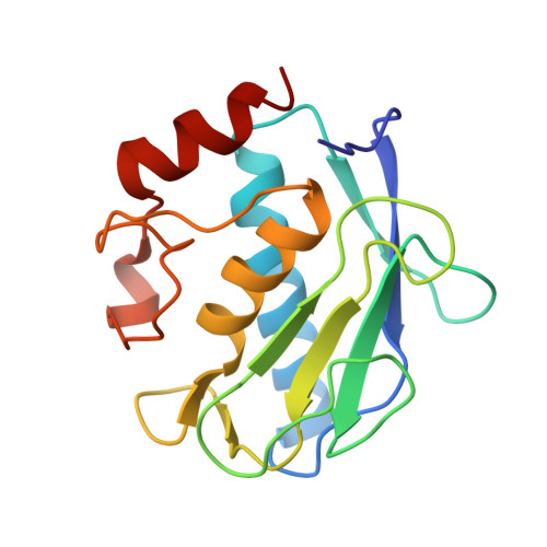
3F1A

Crystal structure of the catalytic domain of human MMP12 complexed with the inhibitor N-(2-nitroso-2-oxoethyl)benzenesulfonamide


Experimental Data Snapshot

  • Method: X-RAY DIFFRACTION
  • Resolution: 1.25 Å
  • R-Value Free: 0.189 
  • R-Value Work: 0.167 
  • R-Value Observed: 0.169 

wwPDB Validation   3D Report Full Report


Ligand Structure Quality Assessment 


This is version 1.3 of the entry. See complete history


Literature

Exploring the subtleties of drug-receptor interactions: the case of matrix metalloproteinases.

Bertini, I.Calderone, V.Fragai, M.Giachetti, A.Loconte, M.Luchinat, C.Maletta, M.Nativi, C.Yeo, K.J.

(2007) J Am Chem Soc 129: 2466-2475

  • DOI: https://doi.org/10.1021/ja065156z
  • Primary Citation of Related Structures:  
    3F15, 3F16, 3F17, 3F18, 3F19, 3F1A, 3LK8, 3NX7

  • PubMed Abstract: 

    By solving high-resolution crystal structures of a large number (14 in this case) of adducts of matrix metalloproteinase 12 (MMP12) with strong, nanomolar, inhibitors all derived from a single ligand scaffold, it is shown that the energetics of the ligand-protein interactions can be accounted for directly from the structures to a level of detail that allows us to rationalize for the differential binding affinity between pairs of closely related ligands. In each case, variations in binding affinities can be traced back to slight improvements or worsening of specific interactions with the protein of one or more ligand atoms. Isothermal calorimetry measurements show that the binding of this class of MMP inhibitors is largely enthalpy driven, but a favorable entropic contribution is always present. The binding enthalpy of acetohydroxamic acid (AHA), the prototype zinc-binding group in MMP drug discovery, has been also accurately measured. In principle, this research permits the planning of either improved inhibitors, or inhibitors with improved selectivity for one or another MMP. The present analysis is applicable to any drug target for which structural information on adducts with a series of homologous ligands can be obtained, while structural information obtained from in silico docking is probably not accurate enough for this type of study.


  • Organizational Affiliation

    Magnetic Resonance Center (CERM), University of Florence, Via L. Sacconi 6, 50019 Sesto Fiorentino, Italy. ivanobertini@cerm.unifi.it


Macromolecules
Find similar proteins by:  (by identity cutoff)  |  3D Structure
Entity ID: 1
MoleculeChains Sequence LengthOrganismDetailsImage
Macrophage metalloelastase158Homo sapiensMutation(s): 1 
Gene Names: HMEMMP12
EC: 3.4.24.65
UniProt & NIH Common Fund Data Resources
Find proteins for P39900 (Homo sapiens)
Explore P39900 
Go to UniProtKB:  P39900
PHAROS:  P39900
GTEx:  ENSG00000262406 
Entity Groups  
Sequence Clusters30% Identity50% Identity70% Identity90% Identity95% Identity100% Identity
UniProt GroupP39900
Sequence Annotations
Expand
  • Reference Sequence
Small Molecules
Binding Affinity Annotations 
IDSourceBinding Affinity
HS7 PDBBind:  3F1A Kd: 61.1 (nM) from 1 assay(s)
Experimental Data & Validation

Experimental Data

  • Method: X-RAY DIFFRACTION
  • Resolution: 1.25 Å
  • R-Value Free: 0.189 
  • R-Value Work: 0.167 
  • R-Value Observed: 0.169 
  • Space Group: C 1 2 1
Unit Cell:
Length ( Å )Angle ( ˚ )
a = 51.834α = 90
b = 60.282β = 115.17
c = 54.178γ = 90
Software Package:
Software NamePurpose
MAR345dtbdata collection
MOLREPphasing
REFMACrefinement
MOSFLMdata reduction
SCALAdata scaling

Structure Validation

View Full Validation Report


Ligand Structure Quality Assessment 


Entry History 

Deposition Data

Revision History  (Full details and data files)

  • Version 1.0: 2008-11-18
    Type: Initial release
  • Version 1.1: 2011-07-13
    Changes: Version format compliance
  • Version 1.2: 2021-11-10
    Changes: Data collection, Database references, Derived calculations
  • Version 1.3: 2023-12-27
    Changes: Data collection