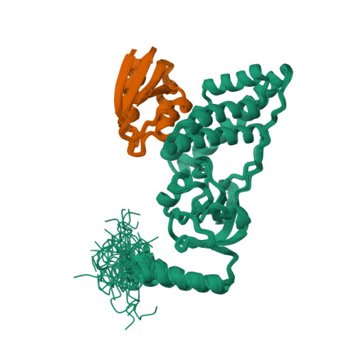
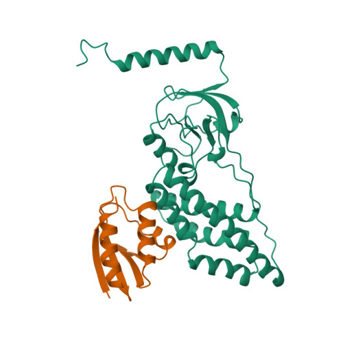
3EZB

COMPLEX OF THE AMINO TERMINAL DOMAIN OF ENZYME I AND THE HISTIDINE-CONTAINING PHOSPHOCARRIER PROTEIN HPR FROM ESCHERICHIA COLI


Experimental Data Snapshot

  • Method: SOLUTION NMR
  • Conformers Calculated: 40 
  • Conformers Submitted: 40 
  • Selection Criteria: REGULARIZED MEAN STRUCTURE 

wwPDB Validation   3D Report Full Report


This is version 1.4 of the entry. See complete history


Literature

Solution structure of the 40,000 Mr phosphoryl transfer complex between the N-terminal domain of enzyme I and HPr.

Garrett, D.S.Seok, Y.J.Peterkofsky, A.Gronenborn, A.M.Clore, G.M.

(1999) Nat Struct Biol 6: 166-173

  • DOI: https://doi.org/10.1038/5854
  • Primary Citation of Related Structures:  
    3EZA, 3EZB, 3EZE

  • PubMed Abstract: 

    The solution structure of the first protein-protein complex of the bacterial phosphoenolpyruvate: sugar phosphotransferase system between the N-terminal domain of enzyme I (EIN) and the histidine-containing phosphocarrier protein HPr has been determined by NMR spectroscopy, including the use of residual dipolar couplings that provide long-range structural information. The complex between EIN and HPr is a classical example of surface complementarity, involving an essentially all helical interface, comprising helices 2, 2', 3 and 4 of the alpha-subdomain of EIN and helices 1 and 2 of HPr, that requires virtually no changes in conformation of the components relative to that in their respective free states. The specificity of the complex is dependent on the correct placement of both van der Waals and electrostatic contacts. The transition state can be formed with minimal changes in overall conformation, and is stabilized in favor of phosphorylated HPr, thereby accounting for the directionality of phosphoryl transfer.


  • Organizational Affiliation

    Laboratory of Chemical Physics, National Institute of Diabetes and Digestive and Kidney Diseases, National Institutes of Health, Bethesda, MD 20892-0520, USA.


Macromolecules
Find similar proteins by:  (by identity cutoff)  |  3D Structure
Entity ID: 1
MoleculeChains Sequence LengthOrganismDetailsImage
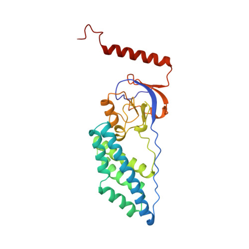
PROTEIN (PHOSPHOTRANSFER SYSTEM, ENZYME I)259Escherichia coliMutation(s): 0 
EC: 2.7.3.9
UniProt
Find proteins for P08839 (Escherichia coli (strain K12))
Explore P08839 
Go to UniProtKB:  P08839
Entity Groups  
Sequence Clusters30% Identity50% Identity70% Identity90% Identity95% Identity100% Identity
UniProt GroupP08839
Sequence Annotations
Expand
  • Reference Sequence
Find similar proteins by:  (by identity cutoff)  |  3D Structure
Entity ID: 2
MoleculeChains Sequence LengthOrganismDetailsImage
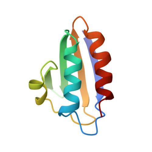
PROTEIN (PHOSPHOCARRIER PROTEIN HPR)85Escherichia coliMutation(s): 0 
UniProt
Find proteins for P0AA04 (Escherichia coli (strain K12))
Explore P0AA04 
Go to UniProtKB:  P0AA04
Entity Groups  
Sequence Clusters30% Identity50% Identity70% Identity90% Identity95% Identity100% Identity
UniProt GroupP0AA04
Sequence Annotations
Expand
  • Reference Sequence
Experimental Data & Validation

Experimental Data

  • Method: SOLUTION NMR
  • Conformers Calculated: 40 
  • Conformers Submitted: 40 
  • Selection Criteria: REGULARIZED MEAN STRUCTURE 

Structure Validation

View Full Validation Report


Entry History 

Deposition Data

Revision History  (Full details and data files)

  • Version 1.0: 1999-12-16
    Type: Initial release
  • Version 1.1: 2008-04-27
    Changes: Version format compliance
  • Version 1.2: 2011-07-13
    Changes: Version format compliance
  • Version 1.3: 2022-03-16
    Changes: Database references, Derived calculations
  • Version 1.4: 2023-12-27
    Changes: Data collection