3EYX | pdb_00003eyx

Crystal structure of Carbonic Anhydrase Nce103 from Saccharomyces cerevisiae


Experimental Data Snapshot

  • Method: X-RAY DIFFRACTION
  • Resolution: 2.04 Å
  • R-Value Free: 
    0.241 (Depositor), 0.241 (DCC) 
  • R-Value Work: 
    0.197 (Depositor), 0.196 (DCC) 
  • R-Value Observed: 
    0.199 (Depositor) 

Starting Model: experimental
View more details

wwPDB Validation 3D Report Full Report

Validation slider image for 3EYX

This is version 1.2 of the entry. See complete history

Literature

Structural insights into the substrate tunnel of Saccharomyces cerevisiae carbonic anhydrase Nce103.

Teng, Y.B.Jiang, Y.L.He, Y.X.He, W.W.Lian, F.M.Chen, Y.Zhou, C.Z.

(2009) BMC Struct Biol 9: 67-67

  • DOI: https://doi.org/10.1186/1472-6807-9-67
  • Primary Citation Related Structures: 
    3EYX

  • PubMed Abstract: 

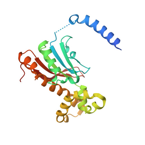
    The carbonic anhydrases (CAs) are involved in inorganic carbon utilization. They have been classified into six evolutionary and structural families: alpha-, beta-, gamma-, delta-, epsilon-, zeta- CAs, with beta-CAs present in higher plants, algae and prokaryotes. The yeast Saccharomyces cerevisiae encodes a single copy of beta-CA Nce103/YNL036W. We determined the crystal structure of Nce103 in complex with a substrate analog at 2.04 A resolution. It assembles as a homodimer, with the active site located at the interface between two monomers. At the bottom of the substrate pocket, a zinc ion is coordinated by the three highly conserved residues Cys57, His112 and Cys115 in addition to a water molecule. Residues Asp59, Arg61, Gly111, Leu102, Val80, Phe75 and Phe97 form a tunnel to the bottom of the active site which is occupied by a molecule of the substrate analog acetate. Activity assays of full length and two truncated versions of Nce103 indicated that the N-terminal arm is indispensable. The quaternary structure of Nce103 resembles the typical plant type beta-CAs of known structure, with an N-terminal arm indispensable for the enzymatic activity. Comparative structure analysis enables us to draw a possible tunnel for the substrate to access the active site which is located at the bottom of a funnel-shaped substrate pocket.


  • Organizational Affiliation
    • Hefei National Laboratory for Physical Sciences at Microscale and School of Life Sciences, University of Science and Technology of China, Hefei, Anhui, 230027, PR China. ybteng@mail.ustc.edu.cn

Macromolecule Content 

  • Total Structure Weight: 49.62 kDa 
  • Atom Count: 3,308 
  • Modeled Residue Count: 394 
  • Deposited Residue Count: 432 
  • Unique protein chains: 1

Macromolecules

Find similar proteins by:|  3D Structure
Entity ID: 1
MoleculeChains  Sequence LengthOrganismDetailsImage
Carbonic anhydrase
A, B
216Saccharomyces cerevisiaeMutation(s): 0 
Gene Names: NCE103
EC: 4.2.1.1
UniProt
Find proteins for P53615 (Saccharomyces cerevisiae (strain ATCC 204508 / S288c))
Explore P53615 
Go to UniProtKB:  P53615
Entity Groups
Sequence Clusters30% Identity50% Identity70% Identity90% Identity95% Identity100% Identity
UniProt GroupP53615
Sequence Annotations
Expand
Reference Sequence

Small Molecules

Ligands 3 Unique
IDChains Name / Formula / InChI Key2D Diagram3D Interactions
ZN

Query on ZN


Download:Ideal Coordinates CCD File
C [auth A],
H [auth B]
ZINC ION
Zn
PTFCDOFLOPIGGS-UHFFFAOYSA-N
EDO

Query on EDO


Download:Ideal Coordinates CCD File
F [auth A],
G [auth A],
I [auth B],
J [auth B]
1,2-ETHANEDIOL
C2 H6 O2
LYCAIKOWRPUZTN-UHFFFAOYSA-N
ACT

Query on ACT


Download:Ideal Coordinates CCD File
D [auth A],
E [auth A]
ACETATE ION
C2 H3 O2
QTBSBXVTEAMEQO-UHFFFAOYSA-M

Experimental Data & Validation

Experimental Data

  • Method: X-RAY DIFFRACTION
  • Resolution: 2.04 Å
  • R-Value Free:  0.241 (Depositor), 0.241 (DCC) 
  • R-Value Work:  0.197 (Depositor), 0.196 (DCC) 
  • R-Value Observed: 0.199 (Depositor) 
Space Group: C 2 2 21
Unit Cell:
Length ( Å )Angle ( ˚ )
a = 60.57α = 90
b = 155.73β = 90
c = 89.69γ = 90
Software Package:
Software NamePurpose
MAR345dtbdata collection
MOLREPphasing
REFMACrefinement
MOSFLMdata reduction
SCALAdata scaling

Structure Validation

View Full Validation Report


Entry History 

Deposition Data

Revision History  (Full details and data files)

  • Version 1.0: 2009-09-15
    Type: Initial release
  • Version 1.1: 2011-07-13
    Changes: Version format compliance
  • Version 1.2: 2023-11-01
    Changes: Data collection, Database references, Derived calculations, Refinement description