3ET9 | pdb_00003et9

Crystal structure of the engineered neutralizing antibody 1H


Experimental Data Snapshot

  • Method: X-RAY DIFFRACTION
  • Resolution: 2.80 Å
  • R-Value Free: 
    0.255 (Depositor), 0.211 (DCC) 
  • R-Value Work: 
    0.217 (Depositor), 0.220 (DCC) 
  • R-Value Observed: 
    0.217 (Depositor) 

Starting Model: experimental
View more details

wwPDB Validation 3D Report Full Report

Validation slider image for 3ET9

This is version 1.6 of the entry. See complete history

Literature

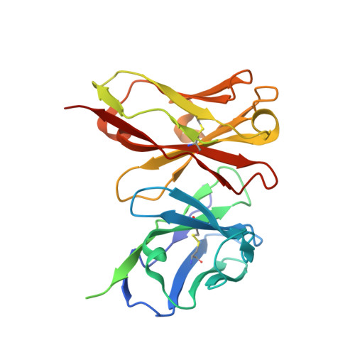
Crystal structure of the engineered neutralizing antibody m18 complexed to domain 4 of the anthrax protective antigen.

Leysath, C.E.Monzingo, A.F.Maynard, J.A.Barnett, J.Georgiou, G.Iverson, B.L.Robertus, J.D.

(2009) J Mol Biology 387: 680-693

  • DOI: https://doi.org/10.1016/j.jmb.2009.02.003
  • Primary Citation Related Structures: 
    3ESU, 3ESV, 3ET9, 3ETB

  • PubMed Abstract: 

    The virulence of Bacillus anthracis is critically dependent on the cytotoxic components of the anthrax toxin, lethal factor (LF) and edema factor (EF). LF and EF gain entry into host cells through interactions with the protective antigen (PA), which binds to host cellular receptors such as CMG2. Antibodies that neutralize PA have been shown to confer protection in animal models and are undergoing intense clinical development. A murine monoclonal antibody, 14B7, has been reported to interact with domain 4 of PA (PAD4) and block its binding to CMG2. More recently, the 14B7 antibody was used as the platform for the selection of very high affinity, single-chain antibodies that have tremendous potential as a combination anthrax prophylactic and treatment. Here, we report the high-resolution X-ray structures of three high-affinity, single-chain antibodies in the 14B7 family; 14B7 and two high-affinity variants 1H and M18. In addition, we present the first neutralizing antibody-PA structure, M18 in complex with PAD4 at 3.8 A resolution. These structures provide insights into the mechanism of neutralization, and the effect of various mutations on antibody affinity, and enable a comparison between the binding of the M18 antibody and CMG2 with PAD4.


  • Organizational Affiliation
    • Institute for Cellular and Molecular Biology, 1 University Station, University of Texas at Austin, Austin, TX 78712, USA.

Macromolecule Content 

  • Total Structure Weight: 26.76 kDa 
  • Atom Count: 1,740 
  • Modeled Residue Count: 227 
  • Deposited Residue Count: 252 
  • Unique protein chains: 1

Macromolecules

Find similar proteins by:|  3D Structure
Entity ID: 1
MoleculeChains  Sequence LengthOrganismDetailsImage
Antibody 1H light chain and antibody 1H heavy chain linked with a synthetic (GGGGS)4 linkerA [auth F]252Mus musculusMutation(s): 3 

Experimental Data & Validation

Experimental Data

  • Method: X-RAY DIFFRACTION
  • Resolution: 2.80 Å
  • R-Value Free:  0.255 (Depositor), 0.211 (DCC) 
  • R-Value Work:  0.217 (Depositor), 0.220 (DCC) 
  • R-Value Observed: 0.217 (Depositor) 
Space Group: P 41 21 2
Unit Cell:
Length ( Å )Angle ( ˚ )
a = 79.94α = 90
b = 79.94β = 90
c = 68.17γ = 90
Software Package:
Software NamePurpose
DENZOdata reduction
SCALEPACKdata scaling
CNSrefinement
PDB_EXTRACTdata extraction
CrystalCleardata collection
HKL-2000data reduction
MOLREPphasing

Structure Validation

View Full Validation Report


Entry History 

Deposition Data

Revision History  (Full details and data files)

  • Version 1.0: 2009-04-28
    Type: Initial release
  • Version 1.1: 2011-07-13
    Changes: Version format compliance
  • Version 1.2: 2017-08-02
    Changes: Source and taxonomy
  • Version 1.3: 2017-10-25
    Changes: Refinement description
  • Version 1.4: 2021-10-20
    Changes: Database references
  • Version 1.5: 2023-09-06
    Changes: Data collection, Refinement description
  • Version 1.6: 2024-11-06
    Changes: Structure summary