3ERR

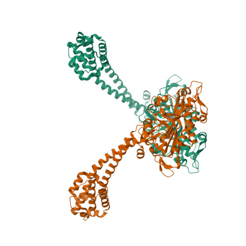
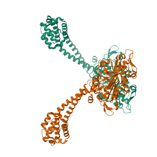
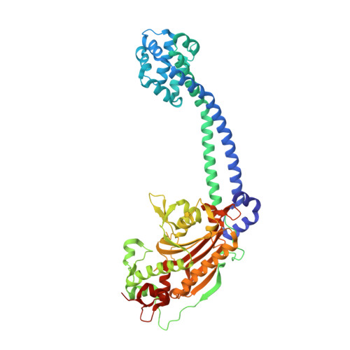
Microtubule binding domain from mouse cytoplasmic dynein as a fusion with seryl-tRNA synthetase


Experimental Data Snapshot

  • Method: X-RAY DIFFRACTION
  • Resolution: 2.27 Å
  • R-Value Free: 0.247 
  • R-Value Work: 0.197 
  • R-Value Observed: 0.200 

Starting Model: experimental
View more details

wwPDB Validation   3D Report Full Report


Ligand Structure Quality Assessment 


This is version 1.4 of the entry. See complete history


Literature

Structure and functional role of dynein's microtubule-binding domain.

Carter, A.P.Garbarino, J.E.Wilson-Kubalek, E.M.Shipley, W.E.Cho, C.Milligan, R.A.Vale, R.D.Gibbons, I.R.

(2008) Science 322: 1691-1695

  • DOI: https://doi.org/10.1126/science.1164424
  • Primary Citation of Related Structures:  
    3ERR

  • PubMed Abstract: 

    Dynein motors move various cargos along microtubules within the cytoplasm and power the beating of cilia and flagella. An unusual feature of dynein is that its microtubule-binding domain (MTBD) is separated from its ring-shaped AAA+ adenosine triphosphatase (ATPase) domain by a 15-nanometer coiled-coil stalk. We report the crystal structure of the mouse cytoplasmic dynein MTBD and a portion of the coiled coil, which supports a mechanism by which the ATPase domain and MTBD may communicate through a shift in the heptad registry of the coiled coil. Surprisingly, functional data suggest that the MTBD, and not the ATPase domain, is the main determinant of the direction of dynein motility.


  • Organizational Affiliation

    Howard Hughes Medical Institute and Department of Cellular and Molecular Pharmacology, University of California, San Francisco, CA 94158, USA.


Macromolecules
Find similar proteins by:  (by identity cutoff)  |  3D Structure
Entity ID: 1
MoleculeChains Sequence LengthOrganismDetailsImage
fusion protein of microtubule binding domain from mouse cytoplasmic dynein and seryl-tRNA synthetase from Thermus thermophilus
A, B
536Mus musculusThermus thermophilus HB27
This entity is chimeric
Mutation(s): 2 
EC: 6.1.1.11
UniProt & NIH Common Fund Data Resources
Find proteins for Q5SJX7 (Thermus thermophilus (strain ATCC 27634 / DSM 579 / HB8))
Explore Q5SJX7 
Go to UniProtKB:  Q5SJX7
Find proteins for Q9JHU4 (Mus musculus)
Explore Q9JHU4 
Go to UniProtKB:  Q9JHU4
IMPC:  MGI:103147
Entity Groups  
Sequence Clusters30% Identity50% Identity70% Identity90% Identity95% Identity100% Identity
UniProt GroupsQ9JHU4Q5SJX7
Sequence Annotations
Expand
  • Reference Sequence
Experimental Data & Validation

Experimental Data

  • Method: X-RAY DIFFRACTION
  • Resolution: 2.27 Å
  • R-Value Free: 0.247 
  • R-Value Work: 0.197 
  • R-Value Observed: 0.200 
  • Space Group: H 3
Unit Cell:
Length ( Å )Angle ( ˚ )
a = 144.302α = 90
b = 144.302β = 90
c = 159.946γ = 120
Software Package:
Software NamePurpose
HKL-2000data collection
PHASERphasing
REFMACrefinement
HKL-2000data reduction
HKL-2000data scaling

Structure Validation

View Full Validation Report


Ligand Structure Quality Assessment 


Entry History 

Deposition Data

  • Released Date: 2008-11-25 
  • Deposition Author(s): Carter, A.P.

Revision History  (Full details and data files)

  • Version 1.0: 2008-11-25
    Type: Initial release
  • Version 1.1: 2011-07-13
    Changes: Version format compliance
  • Version 1.2: 2017-08-02
    Changes: Refinement description, Source and taxonomy
  • Version 1.3: 2021-10-20
    Changes: Database references
  • Version 1.4: 2023-09-06
    Changes: Data collection, Refinement description