3ER5 | pdb_00003er5

THE ACTIVE SITE OF ASPARTIC PROTEINASES


Experimental Data Snapshot

  • Method: X-RAY DIFFRACTION
  • Resolution: 1.80 Å
  • R-Value Work: 
    0.150 (Depositor) 

wwPDB Validation 3D Report Full Report

Validation slider image for 3ER5

This is version 1.5 of the entry. See complete history

Literature

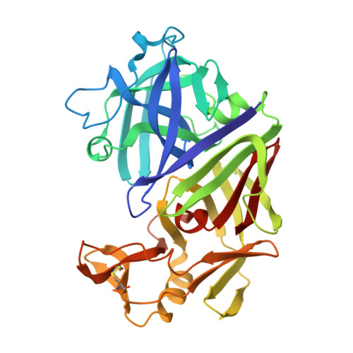
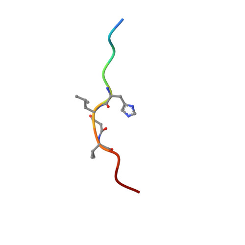
X-ray-crystallographic studies of complexes of pepstatin A and a statine-containing human renin inhibitor with endothiapepsin.

Bailey, D.Cooper, J.B.Veerapandian, B.Blundell, T.L.Atrash, B.Jones, D.M.Szelke, M.

(1993) Biochem J 289 ( Pt 2): 363-371

  • DOI: https://doi.org/10.1042/bj2890363
  • Primary Citation Related Structures: 
    3ER5

  • PubMed Abstract: 

    H-189, a synthetic human renin inhibitor, and pepstatin A, a naturally occurring inhibitor of aspartic proteinases, have been co-crystallized with the fungal aspartic proteinase endothiapepsin (EC 3.4.23.6). H-189 [Pro-His-Pro-Phe-His-Sta-(statyl)-Val-Ile-His-Lys] is an analogue of human angiotensinogen. Pepstatin A [Iva(isovaleryl)-Val-Val-Sta-Ala-Sta] is a blocked pentapeptide which inhibits many aspartic proteinases. The structures of the complexes have been determined by X-ray diffraction and refined to crystallographic R-factors of 0.15 and 0.16 at resolutions of 0.18 nm (1.8 A) and 0.2 nm (2.0 A) respectively. H-189 is in an extended conformation, in which the statine residue is a dipeptide analogue of P1 and P'1 as indicated by the conformation and network of contacts and hydrogen bonds. Pepstatin A has an extended conformation to the P'2 alanine residue, but the leucyl side chain of the terminal statine residue binds back into the S'1 subsite, and an inverse gamma-turn occurs between P'1 and P'3. The hydroxy moiety of the statine at P1 in both complexes displaces the solvent molecule that hydrogen-bonds with the catalytic aspartate residues (32 and 215) in the native enzyme. Solvent molecules originally present in the native structure at the active site are displaced on inhibitor binding (12 when pepstatin A binds; 16 when H-189 binds).


  • Organizational Affiliation
    • Department of Crystallography, Birkbeck College, University of London, U.K.

Macromolecule Content 

  • Total Structure Weight: 35.09 kDa 
  • Atom Count: 2,703 
  • Modeled Residue Count: 340 
  • Deposited Residue Count: 340 
  • Unique protein chains: 2

Macromolecules

Find similar proteins by:|  3D Structure
Entity ID: 1
MoleculeChains  Sequence LengthOrganismDetailsImage
ENDOTHIAPEPSINA [auth E]330Cryphonectria parasiticaMutation(s): 0 
EC: 3.4.23.6 (PDB Primary Data), 3.4.23.22 (UniProt)
UniProt
Find proteins for P11838 (Cryphonectria parasitica)
Explore P11838 
Go to UniProtKB:  P11838
Entity Groups
Sequence Clusters30% Identity50% Identity70% Identity90% Identity95% Identity100% Identity
UniProt GroupP11838
Sequence Annotations
Expand
Reference Sequence
Find similar proteins by:  Sequence   |   3D Structure  
Entity ID: 2
MoleculeChains  Sequence LengthOrganismDetailsImage
H-189B [auth I]10Bos taurusMutation(s): 0 
Gene Names: AGT
UniProt
Find proteins for P01017 (Bos taurus)
Explore P01017 
Go to UniProtKB:  P01017
Entity Groups
Sequence Clusters30% Identity50% Identity70% Identity90% Identity95% Identity100% Identity
UniProt GroupP01017
Sequence Annotations
Expand
Reference Sequence

Biologically Interesting Molecules (External Reference) 

1 Unique
Entity ID: 2
IDChains NameType/Class2D Diagram3D Interactions
PRD_000430
Query on PRD_000430
B [auth I]H 189Peptide-like / Inhibitor

Experimental Data & Validation

Experimental Data

  • Method: X-RAY DIFFRACTION
  • Resolution: 1.80 Å
  • R-Value Work:  0.150 (Depositor) 
Space Group: P 1 21 1
Unit Cell:
Length ( Å )Angle ( ˚ )
a = 43.1α = 90
b = 75.4β = 97
c = 42.8γ = 90
Software Package:
Software NamePurpose
PROLSQrefinement

Structure Validation

View Full Validation Report


Entry History 

Deposition Data

Revision History  (Full details and data files)

  • Version 1.0: 1991-04-15
    Type: Initial release
  • Version 1.1: 2008-03-03
    Changes: Version format compliance
  • Version 1.2: 2011-07-13
    Changes: Atomic model, Database references, Derived calculations, Non-polymer description, Structure summary, Version format compliance
  • Version 1.3: 2012-12-12
    Changes: Other
  • Version 1.4: 2017-11-29
    Changes: Derived calculations, Other
  • Version 1.5: 2024-10-23
    Changes: Data collection, Database references, Derived calculations, Structure summary