3EQS | pdb_00003eqs

Crystal structure of human MDM2 in complex with a 12-mer peptide inhibitor


Experimental Data Snapshot

  • Method: X-RAY DIFFRACTION
  • Resolution: 1.65 Å
  • R-Value Free: 
    0.193 (Depositor), 0.201 (DCC) 
  • R-Value Work: 
    0.154 (Depositor), 0.165 (DCC) 
  • R-Value Observed: 
    0.156 (Depositor) 

Starting Model: experimental
View more details

wwPDB Validation 3D Report Full Report

Validation slider image for 3EQS

This is version 1.2 of the entry. See complete history

Literature

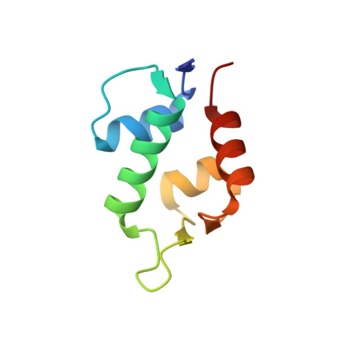
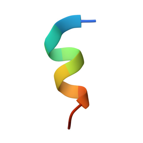
Structural basis for high-affinity peptide inhibition of p53 interactions with MDM2 and MDMX.

Pazgier, M.Liu, M.Zou, G.Yuan, W.Li, C.Li, C.Li, J.Monbo, J.Zella, D.Tarasov, S.G.Lu, W.

(2009) Proc Natl Acad Sci U S A 106: 4665-4670

  • DOI: https://doi.org/10.1073/pnas.0900947106
  • Primary Citation Related Structures: 
    3EQS, 3EQY

  • PubMed Abstract: 

    The oncoproteins MDM2 and MDMX negatively regulate the activity and stability of the tumor suppressor protein p53--a cellular process initiated by MDM2 and/or MDMX binding to the N-terminal transactivation domain of p53. MDM2 and MDMX in many tumors confer p53 inactivation and tumor survival, and are important molecular targets for anticancer therapy. We screened a duodecimal peptide phage library against site-specifically biotinylated p53-binding domains of human MDM2 and MDMX chemically synthesized via native chemical ligation, and identified several peptide inhibitors of the p53-MDM2/MDMX interactions. The most potent inhibitor (TSFAEYWNLLSP), termed PMI, bound to MDM2 and MDMX at low nanomolar affinities--approximately 2 orders of magnitude stronger than the wild-type p53 peptide of the same length (ETFSDLWKLLPE). We solved the crystal structures of synthetic MDM2 and MDMX, both in complex with PMI, at 1.6 A resolution. Comparative structural analysis identified an extensive, tightened intramolecular H-bonding network in bound PMI that contributed to its conformational stability, thus enhanced binding to the 2 oncogenic proteins. Importantly, the C-terminal residue Pro of PMI induced formation of a hydrophobic cleft in MDMX previously unseen in the structures of p53-bound MDM2 or MDMX. Our findings deciphered the structural basis for high-affinity peptide inhibition of p53 interactions with MDM2 and MDMX, shedding new light on structure-based rational design of different classes of p53 activators for potential therapeutic use.


  • Organizational Affiliation
    • Institute of Human Virology, University of Maryland School of Medicine, 725 West Lombard Street, Baltimore, MD 21201, USA.

Macromolecule Content 

  • Total Structure Weight: 11.53 kDa 
  • Atom Count: 1,001 
  • Modeled Residue Count: 95 
  • Deposited Residue Count: 97 
  • Unique protein chains: 2

Macromolecules

Find similar proteins by:|  3D Structure
Entity ID: 1
MoleculeChains  Sequence LengthOrganismDetailsImage
E3 ubiquitin-protein ligase Mdm285N/AMutation(s): 0 
EC: 6.3.2 (PDB Primary Data), 2.3.2.27 (UniProt)
UniProt & NIH Common Fund Data Resources
Find proteins for Q00987 (Homo sapiens)
Explore Q00987 
Go to UniProtKB:  Q00987
PHAROS:  Q00987
GTEx:  ENSG00000135679 
Entity Groups
Sequence Clusters30% Identity50% Identity70% Identity90% Identity95% Identity100% Identity
UniProt GroupQ00987
Sequence Annotations
Expand
Reference Sequence
Find similar proteins by:  Sequence   |   3D Structure  
Entity ID: 2
MoleculeChains  Sequence LengthOrganismDetailsImage
12-mer peptide inhibitor12N/AMutation(s): 0 

Small Molecules

Ligands 1 Unique
IDChains Name / Formula / InChI Key2D Diagram3D Interactions
GAI

Query on GAI


Download:Ideal Coordinates CCD File
C [auth A]GUANIDINE
C H5 N3
ZRALSGWEFCBTJO-UHFFFAOYSA-N

Experimental Data & Validation

Experimental Data

  • Method: X-RAY DIFFRACTION
  • Resolution: 1.65 Å
  • R-Value Free:  0.193 (Depositor), 0.201 (DCC) 
  • R-Value Work:  0.154 (Depositor), 0.165 (DCC) 
  • R-Value Observed: 0.156 (Depositor) 
Space Group: C 1 2 1
Unit Cell:
Length ( Å )Angle ( ˚ )
a = 71.517α = 90
b = 43.274β = 116.51
c = 35.036γ = 90
Software Package:
Software NamePurpose
HKL-2000data collection
PHASERphasing
REFMACrefinement
DENZOdata reduction
SCALEPACKdata scaling

Structure Validation

View Full Validation Report


Entry History 

Deposition Data

Revision History  (Full details and data files)

  • Version 1.0: 2009-03-17
    Type: Initial release
  • Version 1.1: 2011-07-13
    Changes: Advisory, Version format compliance
  • Version 1.2: 2023-09-06
    Changes: Data collection, Database references, Derived calculations, Refinement description