3ENG | pdb_00003eng

STRUCTURE OF ENDOGLUCANASE V CELLOBIOSE COMPLEX


Experimental Data Snapshot

  • Method: X-RAY DIFFRACTION
  • Resolution: 1.90 Å
  • R-Value Work: 
    0.145 (Depositor) 

Starting Model: experimental
View more details

wwPDB Validation 3D Report Full Report

Validation slider image for 3ENG

This is version 2.2 of the entry. See complete history

Literature

Structure determination and refinement of the Humicola insolens endoglucanase V at 1.5 A resolution.

Davies, G.J.Dodson, G.Moore, M.H.Tolley, S.P.Dauter, Z.Wilson, K.S.Rasmussen, G.Schulein, M.

(1996) Acta Crystallogr D Biol Crystallogr 52: 7-17

  • DOI: https://doi.org/10.1107/S0907444995009280
  • Primary Citation Related Structures: 
    3ENG, 4ENG

  • PubMed Abstract: 

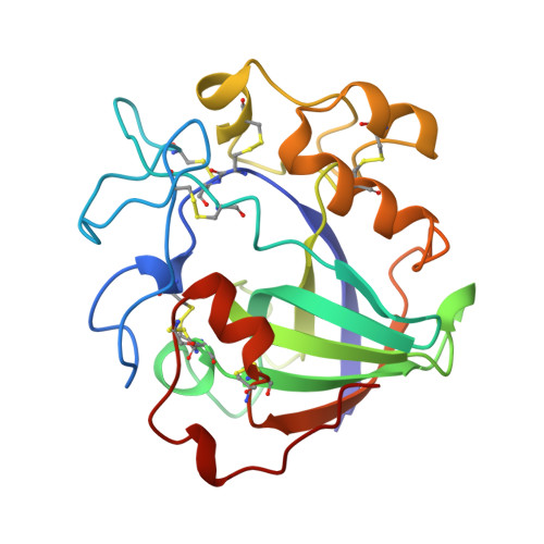
    The structure of the catalytic core of the endoglucanase V (EGV) from Humicola insolens has been determined by the method of multiple isomorphous replacement at 1.5 A resolution. The final model, refined with X-PLOR and PROLSQ, has a crystallographic R factor of 0.163 (R(free) = 0.240) with deviations from stereochemical target values of 0.012 A and 0.037 degrees for bonds and angles, respectively. The model was further refined with SHELXL, including anisotropic modelling of the protein-atom temperature factors, to give a final model with an R factor of 0.105 and an R(free) of 0.154. The initial isomorphous replacement electron-density map was poor and uninterpretable but was improved by the use of synchrotron data collected at a wavelength chosen so as to optimize the f" contribution of the anomalous scattering from the heavy atoms. The structure of H. insolens EGV consists of a six-stranded beta-barrel domain, similar to that found in a family of plant defence proteins, linked by a number of disulfide-bonded loop regions. A long open groove runs across the surface of the enzyme either side of which lie the catalytic aspartate residues. The 9 A separation of the catalytic carboxylate groups is consistent with the observation that EGV catalyzes the hydrolysis of the cellulose, beta(1-->4) links with inversion of configuration at the anomeric C1 atom. This structure is the first representative from the glycosyl hydrolase family 45.


  • Organizational Affiliation
    • Department of Chemistry, University of York, Heslington, England.

Macromolecule Content 

  • Total Structure Weight: 23.23 kDa 
  • Atom Count: 1,773 
  • Modeled Residue Count: 213 
  • Deposited Residue Count: 213 
  • Unique protein chains: 1

Macromolecules

Find similar proteins by:|  3D Structure
Entity ID: 1
MoleculeChains  Sequence LengthOrganismDetailsImage
ENDOGLUCANASE V CELLOBIOSE COMPLEX213Mycothermus thermophilusMutation(s): 0 
EC: 3.2.1.4
UniProt
Find proteins for P43316 (Humicola insolens)
Explore P43316 
Go to UniProtKB:  P43316
Entity Groups
Sequence Clusters30% Identity50% Identity70% Identity90% Identity95% Identity100% Identity
UniProt GroupP43316
Sequence Annotations
Expand
Reference Sequence

Oligosaccharides

Help  
Entity ID: 2
MoleculeChains Length2D Diagram GlycosylationD Interactions
beta-D-glucopyranose-(1-4)-beta-D-glucopyranose
B
2N/A
Glycosylation Resources
GlyTouCan: G84824ZO
GlyCosmos: G84824ZO

Biologically Interesting Molecules (External Reference) 

1 Unique
Entity ID: 2
IDChains NameType/Class2D Diagram3D Interactions
PRD_900005
Query on PRD_900005
B
beta-cellobioseOligosaccharide / Metabolism

Experimental Data & Validation

Experimental Data

  • Method: X-RAY DIFFRACTION
  • Resolution: 1.90 Å
  • R-Value Work:  0.145 (Depositor) 
Space Group: P 1 21 1
Unit Cell:
Length ( Å )Angle ( ˚ )
a = 41.75α = 90
b = 56.18β = 93.4
c = 37.37γ = 90
Software Package:
Software NamePurpose
DENZOdata reduction
ROTAVATAdata reduction
AMoREphasing
REFMACrefinement
CCP4data scaling

Structure Validation

View Full Validation Report


Entry History 

Deposition Data

Revision History  (Full details and data files)

  • Version 1.0: 1997-06-16
    Type: Initial release
  • Version 1.1: 2008-03-03
    Changes: Version format compliance
  • Version 1.2: 2011-07-13
    Changes: Version format compliance
  • Version 1.3: 2018-05-02
    Changes: Data collection
  • Version 2.0: 2020-07-29
    Type: Remediation
    Reason: Carbohydrate remediation
    Changes: Atomic model, Data collection, Derived calculations, Non-polymer description, Structure summary
  • Version 2.1: 2023-08-09
    Changes: Database references, Refinement description, Structure summary
  • Version 2.2: 2024-10-16
    Changes: Data collection, Structure summary