3EIV | pdb_00003eiv

Crystal Structure of Single-stranded DNA-binding protein from Streptomyces coelicolor


Experimental Data Snapshot

  • Method: X-RAY DIFFRACTION
  • Resolution: 2.14 Å
  • R-Value Free: 
    0.259 (Depositor), 0.226 (DCC) 
  • R-Value Work: 
    0.230 (Depositor), 0.228 (DCC) 
  • R-Value Observed: 
    0.232 (Depositor) 

Starting Model: experimental
View more details

wwPDB Validation 3D Report Full Report

Validation slider image for 3EIV

This is version 1.2 of the entry. See complete history

Literature

Structure of the single-stranded DNA-binding protein from Streptomyces coelicolor.

Stefanic, Z.Vujaklija, D.Luic, M.

(2009) Acta Crystallogr D Biol Crystallogr 65: 974-979

  • DOI: https://doi.org/10.1107/S0907444909023634
  • Primary Citation Related Structures: 
    3EIV

  • PubMed Abstract: 

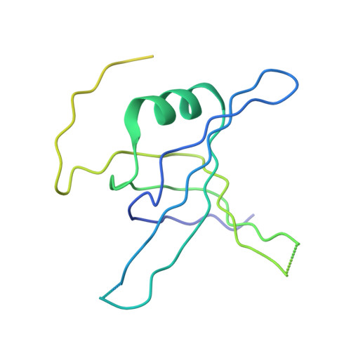
    The crystal structure of the single-stranded DNA-binding protein (SSB) from Streptomyces coelicolor, a filamentous soil bacterium with a complex life cycle and a linear chromosome, has been solved and refined at 2.1 A resolution. The three-dimensional structure shows a common conserved central OB-fold that is found in all structurally determined SSB proteins. However, it shows variations in quaternary structure that have previously only been found in mycobacterial SSBs. The strand involved in the clamp mechanism characteristic of this type of quaternary structure leads to higher stability of the homotetramer. To the best of our knowledge, this is the first X-ray structure of an SSB protein from a member of the genus Streptomyces and it was predicted to be the most stable of the structurally characterized bacterial or human mitochondrial SSBs.


  • Organizational Affiliation
    • Rudjer Bosković Institute, Bijenicka cesta 54, Zagreb, Croatia.

Macromolecule Content 

  • Total Structure Weight: 79.7 kDa 
  • Atom Count: 3,434 
  • Modeled Residue Count: 420 
  • Deposited Residue Count: 796 
  • Unique protein chains: 1

Macromolecules

Find similar proteins by:|  3D Structure
Entity ID: 1
MoleculeChains  Sequence LengthOrganismDetailsImage
Single-stranded DNA-binding protein 2
A, B, C, D
199Streptomyces coelicolorMutation(s): 0 
UniProt
Find proteins for Q9X8U3 (Streptomyces coelicolor (strain ATCC BAA-471 / A3(2) / M145))
Explore Q9X8U3 
Go to UniProtKB:  Q9X8U3
Entity Groups
Sequence Clusters30% Identity50% Identity70% Identity90% Identity95% Identity100% Identity
UniProt GroupQ9X8U3
Sequence Annotations
Expand
Reference Sequence

Experimental Data & Validation

Experimental Data

  • Method: X-RAY DIFFRACTION
  • Resolution: 2.14 Å
  • R-Value Free:  0.259 (Depositor), 0.226 (DCC) 
  • R-Value Work:  0.230 (Depositor), 0.228 (DCC) 
  • R-Value Observed: 0.232 (Depositor) 
Space Group: I 2 2 2
Unit Cell:
Length ( Å )Angle ( ˚ )
a = 101.454α = 90
b = 104.788β = 90
c = 163.306γ = 90
Software Package:
Software NamePurpose
PHASERphasing
PHENIXrefinement
PDB_EXTRACTdata extraction
HKL-3000data collection
X-GENdata reduction
X-GENdata scaling

Structure Validation

View Full Validation Report


Entry History 

Deposition Data

Revision History  (Full details and data files)

  • Version 1.0: 2009-09-08
    Type: Initial release
  • Version 1.1: 2011-07-13
    Changes: Version format compliance
  • Version 1.2: 2023-08-30
    Changes: Data collection, Database references, Refinement description