Mechanism of substrate recognition and PLP-induced conformational changes in LL-diaminopimelate aminotransferase from Arabidopsis thaliana.
Watanabe, N., Clay, M.D., van Belkum, M.J., Cherney, M.M., Vederas, J.C., James, M.N.(2008) J Mol Biol 384: 1314-1329
- PubMed: 18952095 Search on PubMed
- DOI: https://doi.org/10.1016/j.jmb.2008.10.022
- Primary Citation Related Structures:
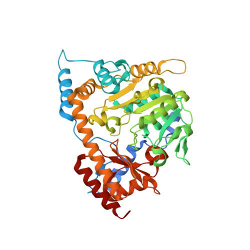
3EI5, 3EI6, 3EI7, 3EI8, 3EI9, 3EIA, 3EIB - PubMed Abstract:
LL-Diaminopimelate aminotransferase (LL-DAP-AT), a pyridoxal phosphate (PLP)-dependent enzyme in the lysine biosynthetic pathways of plants and Chlamydia, is a potential target for the development of herbicides or antibiotics. This homodimeric enzyme converts L-tetrahydrodipicolinic acid (THDP) directly to LL-DAP using L-glutamate as the source of the amino group. Earlier, we described the 3D structures of native and malate-bound LL-DAP-AT from Arabidopsis thaliana (AtDAP-AT). Seven additional crystal structures of AtDAP-AT and its variants are reported here as part of an investigation into the mechanism of substrate recognition and catalysis. Two structures are of AtDAP-AT with reduced external aldimine analogues: N-(5'-phosphopyridoxyl)-L-glutamate (PLP-Glu) and N-(5'-phosphopyridoxyl)- LL-Diaminopimelate (PLP-DAP) bound in the active site. Surprisingly, they reveal that both L-glutamate and LL-DAP are recognized in a very similar fashion by the same sets of amino acid residues; both molecules adopt twisted V-shaped conformations. With both substrates, the alpha-carboxylates are bound in a salt bridge with Arg404, whereas the distal carboxylates are recognized via hydrogen bonds to the well-conserved side chains of Tyr37, Tyr125 and Lys129. The distal C(epsilon) amino group of LL-DAP is specifically recognized by several non-covalent interactions with residues from the other subunit (Asn309*, Tyr94*, Gly95*, and Glu97* (Amino acid designators followed by an asterisk (*) indicate that the residues originate in the other subunit of the dimer)) and by three bound water molecules. Two catalytically inactive variants of AtDAP-AT were created via site-directed mutagenesis of the active site lysine (K270N and K270Q). The structures of these variants permitted the observation of the unreduced external aldimines of PLP with L-glutamate and with LL-DAP in the active site, and revealed differences in the torsion angle about the PLP-substrate bond. Lastly, an apo-AtDAP-AT structure missing PLP revealed details of conformational changes induced by PLP binding and substrate entry into the active site.
- Department of Biochemistry, University of Alberta, Edmonton, Alberta, Canada T6G 2H7.
Organizational Affiliation: