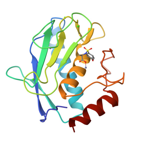
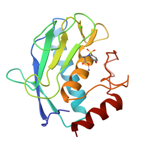
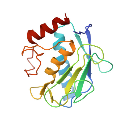
3EHY

Crystal structure of the catalytic domain of human MMP12 complexed with the inhibitor (R)-2-(4-methoxyphenylsulfonamido)propanoic acid


Experimental Data Snapshot

  • Method: X-RAY DIFFRACTION
  • Resolution: 1.90 Å
  • R-Value Free: 0.204 
  • R-Value Work: 0.155 
  • R-Value Observed: 0.159 

Starting Model: experimental
View more details

wwPDB Validation   3D Report Full Report


Ligand Structure Quality Assessment 


This is version 1.3 of the entry. See complete history


Literature

Biotin-tagged probes for MMP expression and activation: design, synthesis, and binding properties

Dragoni, E.Calderone, V.Fragai, M.Jaiswal, R.Luchinat, C.Nativi, C.

(2009) Bioconjug Chem 20: 719-727

  • DOI: https://doi.org/10.1021/bc8003827
  • Primary Citation of Related Structures:  
    3EHX, 3EHY

  • PubMed Abstract: 

    The design and synthesis of biotin chain-terminated inhibitors (BTI) showing high affinity for matrix metalloproteinases (MMPs) on one side and high affinity for avidin through the biotinylated tag on the other are reported. The affinity of the designed BTI toward five different MMPs has been evaluated and the simultaneous formation of a highly stable ternary system Avidin-BTI-MMP clearly assessed. This system will permit the development of new approaches to detect, quantify, or collect MMPs in biological samples, with potential applications in vivo.


  • Organizational Affiliation

    Magnetic Resonance Center - University of Florence, Sesto Fiorentino, Italy.


Macromolecules
Find similar proteins by:  (by identity cutoff)  |  3D Structure
Entity ID: 1
MoleculeChains Sequence LengthOrganismDetailsImage
Macrophage metalloelastase158Homo sapiensMutation(s): 1 
Gene Names: MMP12
EC: 3.4.24.65
UniProt & NIH Common Fund Data Resources
Find proteins for P39900 (Homo sapiens)
Explore P39900 
Go to UniProtKB:  P39900
PHAROS:  P39900
GTEx:  ENSG00000262406 
Entity Groups  
Sequence Clusters30% Identity50% Identity70% Identity90% Identity95% Identity100% Identity
UniProt GroupP39900
Sequence Annotations
Expand
  • Reference Sequence
Small Molecules
Binding Affinity Annotations 
IDSourceBinding Affinity
TBL PDBBind:  3EHY Ki: 1400 (nM) from 1 assay(s)
Experimental Data & Validation

Experimental Data

  • Method: X-RAY DIFFRACTION
  • Resolution: 1.90 Å
  • R-Value Free: 0.204 
  • R-Value Work: 0.155 
  • R-Value Observed: 0.159 
  • Space Group: C 1 2 1
Unit Cell:
Length ( Å )Angle ( ˚ )
a = 51.256α = 90
b = 60.479β = 116.17
c = 54.598γ = 90
Software Package:
Software NamePurpose
CrysalisProdata collection
MOLREPphasing
REFMACrefinement
MOSFLMdata reduction
SCALAdata scaling

Structure Validation

View Full Validation Report


Ligand Structure Quality Assessment 


Entry History 

Deposition Data

Revision History  (Full details and data files)

  • Version 1.0: 2009-05-19
    Type: Initial release
  • Version 1.1: 2011-07-13
    Changes: Version format compliance
  • Version 1.2: 2021-11-10
    Changes: Database references, Derived calculations
  • Version 1.3: 2023-11-01
    Changes: Data collection, Refinement description