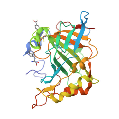
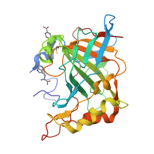
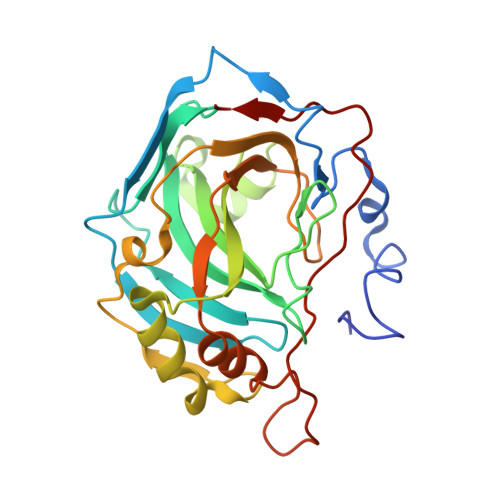
3EFI

Carbonic anhydrase activators: Kinetic and X-ray crystallographic study for the interaction of d- and l-tryptophan with the mammalian isoforms I-XIV


Experimental Data Snapshot

  • Method: X-RAY DIFFRACTION
  • Resolution: 1.75 Å
  • R-Value Free: 0.222 
  • R-Value Work: 0.189 
  • R-Value Observed: 0.191 

Starting Model: experimental
View more details

wwPDB Validation   3D Report Full Report


Ligand Structure Quality Assessment 


This is version 1.3 of the entry. See complete history


Literature

Carbonic anhydrase activators: kinetic and X-ray crystallographic study for the interaction of D- and L-tryptophan with the mammalian isoforms I-XIV

Temperini, C.Innocenti, A.Scozzafava, A.Supuran, C.T.

(2008) Bioorg Med Chem 16: 8373-8378

  • DOI: https://doi.org/10.1016/j.bmc.2008.08.043
  • Primary Citation of Related Structures:  
    3EFI

  • PubMed Abstract: 

    An activation study of mammalian carbonic anhydrase (CA, EC 4.2.1.1) isoforms I-XIV with D- and L-tryptophan has been performed both by means of kinetic and X-ray crystallographic techniques. These compounds show a time dependent activity against isozyme CA II, with activation constants of 1.13 microM for L-Trp and 0.37 microM for D-Trp, respectively, after 24 h of incubation between enzyme and activator. The high resolution X-ray crystal structure of the hCA II-D-Trp adduct revealed the activator to bind in a totally unprecedented way to the enzyme active site as compared to histamine, L-/D-Phe, L-/D-His or L-adrenaline. D-Trp is anchored at the edge of the CA II active site entrance, strongly interacting with amino acid residues Asp130, Phe131 and Gly132 as well as with a loop of a second symmetry related protein molecule from the asymmetric unit, by means of hydrogen bonds and several weak van der Waals interactions involving Glu234, Gly235, Glu236 and Glu238. Thus, a second activator binding site (B) within the CA II cavity has been detected, where only D-Trp was shown so far to bind, in addition to the activator binding site A, in which histamine, L-/D-Phe, and L-/D-His are bound. These findings explain the strong affinity of D-Trp for CA II and may be useful for designing novel classes of CA activators by using this compound as lead molecule.


  • Organizational Affiliation

    Università degli Studi di Firenze, Laboratorio di Chimica Bioinorganica, Room 188, Via della Lastruccia 3, I-50019 Sesto Fiorentino (Firenze), Italy.


Macromolecules
Find similar proteins by:  (by identity cutoff)  |  3D Structure
Entity ID: 1
MoleculeChains Sequence LengthOrganismDetailsImage
Carbonic anhydrase 2260N/AMutation(s): 0 
EC: 4.2.1.1 (PDB Primary Data), 4.2.1.69 (UniProt)
UniProt & NIH Common Fund Data Resources
Find proteins for P00918 (Homo sapiens)
Explore P00918 
Go to UniProtKB:  P00918
PHAROS:  P00918
GTEx:  ENSG00000104267 
Entity Groups  
Sequence Clusters30% Identity50% Identity70% Identity90% Identity95% Identity100% Identity
UniProt GroupP00918
Sequence Annotations
Expand
  • Reference Sequence
Experimental Data & Validation

Experimental Data

  • Method: X-RAY DIFFRACTION
  • Resolution: 1.75 Å
  • R-Value Free: 0.222 
  • R-Value Work: 0.189 
  • R-Value Observed: 0.191 
  • Space Group: P 1 21 1
Unit Cell:
Length ( Å )Angle ( ˚ )
a = 42.07α = 90
b = 41.32β = 104.4
c = 72.05γ = 90
Software Package:
Software NamePurpose
CrysalisProdata collection
AMoREphasing
REFMACrefinement
CrysalisProdata reduction
SCALAdata scaling

Structure Validation

View Full Validation Report


Ligand Structure Quality Assessment 


Entry History 

Deposition Data

Revision History  (Full details and data files)

  • Version 1.0: 2008-09-30
    Type: Initial release
  • Version 1.1: 2011-07-13
    Changes: Version format compliance
  • Version 1.2: 2017-10-25
    Changes: Refinement description
  • Version 1.3: 2023-11-01
    Changes: Data collection, Database references, Derived calculations, Refinement description