3ED3 | pdb_00003ed3

Crystal Structure of the Yeast Dithiol/Disulfide Oxidoreductase Mpd1p


Experimental Data Snapshot

  • Method: X-RAY DIFFRACTION
  • Resolution: 2.00 Å
  • R-Value Free: 
    0.255 (Depositor), 0.256 (DCC) 
  • R-Value Work: 
    0.219 (Depositor), 0.219 (DCC) 

wwPDB Validation 3D Report Full Report

Validation slider image for 3ED3

This is version 1.2 of the entry. See complete history

Literature

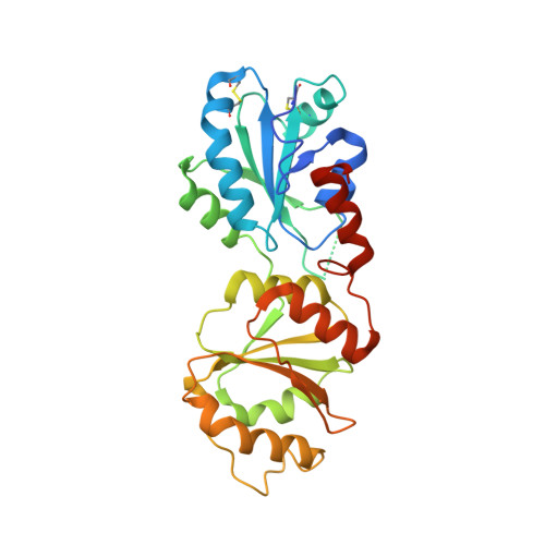
Yeast Mpd1p reveals the structural diversity of the protein disulfide isomerase family

Vitu, E.Gross, E.Greenblatt, H.M.Sevier, C.S.Kaiser, C.A.Fass, D.

(2008) J Mol Biology 384: 631-640

  • DOI: https://doi.org/10.1016/j.jmb.2008.09.052
  • Primary Citation Related Structures: 
    3ED3

  • PubMed Abstract: 

    Oxidoreductases belonging to the protein disulfide isomerase (PDI) family promote proper disulfide bond formation in substrate proteins in the endoplasmic reticulum. In plants and metazoans, new family members continue to be identified and assigned to various functional niches. PDI-like proteins typically contain tandem thioredoxin-fold domains. The limited information available suggested that the relative orientations of these domains may be quite uniform across the family, and structural models based on this assumption are appearing. However, the X-ray crystal structure of the yeast PDI family protein Mpd1p, described here, demonstrates the radically different domain orientations and surface properties achievable with multiple copies of the thioredoxin fold. A comparison of Mpd1p with yeast Pdi1p expands our perspective on the contexts in which redox-active motifs are presented in the PDI family.


  • Organizational Affiliation
    • Department of Structural Biology, Weizmann Institute of Science, Rehovot 76100, Israel.

Macromolecule Content 

  • Total Structure Weight: 69.24 kDa 
  • Atom Count: 4,656 
  • Modeled Residue Count: 533 
  • Deposited Residue Count: 596 
  • Unique protein chains: 1

Macromolecules

Find similar proteins by:|  3D Structure
Entity ID: 1
MoleculeChains  Sequence LengthOrganismDetailsImage
Protein disulfide-isomerase MPD1
A, B
298Saccharomyces cerevisiaeMutation(s): 0 
Gene Names: MPD1
EC: 5.3.4.1
UniProt
Find proteins for Q12404 (Saccharomyces cerevisiae (strain ATCC 204508 / S288c))
Explore Q12404 
Go to UniProtKB:  Q12404
Entity Groups
Sequence Clusters30% Identity50% Identity70% Identity90% Identity95% Identity100% Identity
UniProt GroupQ12404
Sequence Annotations
Expand
Reference Sequence

Small Molecules

Ligands 2 Unique
IDChains Name / Formula / InChI Key2D Diagram3D Interactions
EDO

Query on EDO


Download:Ideal Coordinates CCD File
I [auth A],
J [auth A],
N [auth B],
O [auth B]
1,2-ETHANEDIOL
C2 H6 O2
LYCAIKOWRPUZTN-UHFFFAOYSA-N
ACT

Query on ACT


Download:Ideal Coordinates CCD File
C [auth A]
D [auth A]
E [auth A]
F [auth A]
G [auth A]
C [auth A],
D [auth A],
E [auth A],
F [auth A],
G [auth A],
H [auth A],
K [auth B],
L [auth B],
M [auth B]
ACETATE ION
C2 H3 O2
QTBSBXVTEAMEQO-UHFFFAOYSA-M

Experimental Data & Validation

Experimental Data

  • Method: X-RAY DIFFRACTION
  • Resolution: 2.00 Å
  • R-Value Free:  0.255 (Depositor), 0.256 (DCC) 
  • R-Value Work:  0.219 (Depositor), 0.219 (DCC) 
Space Group: P 1 21 1
Unit Cell:
Length ( Å )Angle ( ˚ )
a = 48.157α = 90
b = 92.268β = 92.23
c = 79.337γ = 90
Software Package:
Software NamePurpose
SOLVEphasing
CNSrefinement
DENZOdata reduction
SCALEPACKdata scaling

Structure Validation

View Full Validation Report


Entry History 

Deposition Data

Revision History  (Full details and data files)

  • Version 1.0: 2008-11-04
    Type: Initial release
  • Version 1.1: 2011-07-13
    Changes: Version format compliance
  • Version 1.2: 2024-11-20
    Changes: Data collection, Database references, Derived calculations, Structure summary