3EAD | pdb_00003ead

Crystal structure of CALX-CBD1


Experimental Data Snapshot

  • Method: X-RAY DIFFRACTION
  • Resolution: 2.25 Å
  • R-Value Free: 
    0.269 (Depositor), 0.310 (DCC) 
  • R-Value Work: 
    0.210 (Depositor), 0.266 (DCC) 
  • R-Value Observed: 
    0.213 (Depositor) 

Starting Model: experimental
View more details

wwPDB Validation 3D Report Full Report

Validation slider image for 3EAD

This is version 1.4 of the entry. See complete history

Literature

Crystal structures of progressive Ca2+ binding states of the Ca2+ sensor Ca2+ binding domain 1 (CBD1) from the CALX Na+/Ca2+ exchanger reveal incremental conformational transitions.

Wu, M.Le, H.D.Wang, M.Yurkov, V.Omelchenko, A.Hnatowich, M.Nix, J.Hryshko, L.V.Zheng, L.

(2010) J Biological Chem 285: 2554-2561

  • DOI: https://doi.org/10.1074/jbc.M109.059162
  • Primary Citation Related Structures: 
    3E9T, 3EAD

  • PubMed Abstract: 

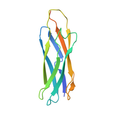
    Na(+)/Ca(2+) exchangers (NCX) constitute a major Ca(2+) export system that facilitates the re-establishment of cytosolic Ca(2+) levels in many tissues. Ca(2+) interactions at its Ca(2+) binding domains (CBD1 and CBD2) are essential for the allosteric regulation of Na(+)/Ca(2+) exchange activity. The structure of the Ca(2+)-bound form of CBD1, the primary Ca(2+) sensor from canine NCX1, but not the Ca(2+)-free form, has been reported, although the molecular mechanism of Ca(2+) regulation remains unclear. Here, we report crystal structures for three distinct Ca(2+) binding states of CBD1 from CALX, a Na(+)/Ca(2+) exchanger found in Drosophila sensory neurons. The fully Ca(2+)-bound CALX-CBD1 structure shows that four Ca(2+) atoms bind at identical Ca(2+) binding sites as those found in NCX1 and that the partial Ca(2+) occupancy and apoform structures exhibit progressive conformational transitions, indicating incremental regulation of CALX exchange by successive Ca(2+) binding at CBD1. The structures also predict that the primary Ca(2+) pair plays the main role in triggering functional conformational changes. Confirming this prediction, mutagenesis of Glu(455), which coordinates the primary Ca(2+) pair, produces dramatic reductions of the regulatory Ca(2+) affinity for exchange current, whereas mutagenesis of Glu(520), which coordinates the secondary Ca(2+) pair, has much smaller effects. Furthermore, our structures indicate that Ca(2+) binding only enhances the stability of the Ca(2+) binding site of CBD1 near the hinge region while the overall structure of CBD1 remains largely unaffected, implying that the Ca(2+) regulatory function of CBD1, and possibly that for the entire NCX family, is mediated through domain interactions between CBD1 and the adjacent CBD2 at this hinge.


  • Organizational Affiliation
    • Center for Membrane Biology, Department of Biochemistry and Molecular Biology, University of Texas Medical School at Houston, Houston, Texas 77030, USA.

Macromolecule Content 

  • Total Structure Weight: 62.66 kDa 
  • Atom Count: 3,693 
  • Modeled Residue Count: 448 
  • Deposited Residue Count: 548 
  • Unique protein chains: 1

Macromolecules

Find similar proteins by:|  3D Structure
Entity ID: 1
MoleculeChains  Sequence LengthOrganismDetailsImage
Na/Ca exchange protein
A, B, C, D
137Drosophila melanogasterMutation(s): 0 
Gene Names: CalxNCXCG5685
UniProt
Find proteins for A0A0B4K6E2 (Drosophila melanogaster)
Explore A0A0B4K6E2 
Go to UniProtKB:  A0A0B4K6E2
Entity Groups
Sequence Clusters30% Identity50% Identity70% Identity90% Identity95% Identity100% Identity
UniProt GroupA0A0B4K6E2
Sequence Annotations
Expand
Reference Sequence

Small Molecules

Ligands 2 Unique
IDChains Name / Formula / InChI Key2D Diagram3D Interactions
GOL

Query on GOL


Download:Ideal Coordinates CCD File
I [auth A]GLYCEROL
C3 H8 O3
PEDCQBHIVMGVHV-UHFFFAOYSA-N
CA

Query on CA


Download:Ideal Coordinates CCD File
E [auth A]
F [auth A]
G [auth A]
H [auth A]
J [auth B]
E [auth A],
F [auth A],
G [auth A],
H [auth A],
J [auth B],
K [auth B],
L [auth B],
M [auth B],
N [auth C],
O [auth C],
P [auth C],
Q [auth C],
R [auth D],
S [auth D],
T [auth D],
U [auth D]
CALCIUM ION
Ca
BHPQYMZQTOCNFJ-UHFFFAOYSA-N

Experimental Data & Validation

Experimental Data

  • Method: X-RAY DIFFRACTION
  • Resolution: 2.25 Å
  • R-Value Free:  0.269 (Depositor), 0.310 (DCC) 
  • R-Value Work:  0.210 (Depositor), 0.266 (DCC) 
  • R-Value Observed: 0.213 (Depositor) 
Space Group: P 21 21 21
Unit Cell:
Length ( Å )Angle ( ˚ )
a = 59.372α = 90
b = 73.722β = 90
c = 129.713γ = 90
Software Package:
Software NamePurpose
MAR345data collection
PHASERphasing
REFMACrefinement
XDSdata reduction
XSCALEdata scaling

Structure Validation

View Full Validation Report


Entry History 

Deposition Data

Revision History  (Full details and data files)

  • Version 1.0: 2009-09-08
    Type: Initial release
  • Version 1.1: 2011-07-13
    Changes: Non-polymer description, Version format compliance
  • Version 1.2: 2017-10-25
    Changes: Refinement description
  • Version 1.3: 2023-07-05
    Changes: Database references, Derived calculations
  • Version 1.4: 2023-09-20
    Changes: Data collection, Refinement description